Quantcast
Channel: Lori-Lynne's Coding Coach Blog
Viewing all 99 articles
Browse latest View live

Article 0

$
0
0
When Coders don’t agree, who is correct?   The Physician office?  The Hospital Facility? 

by Lori-Lynne A. Webb, CPC, CCS-P, CCP, CHDA, COBGC, CDIP

Coding can be a challenge when coders are confronted with procedures that are new, and include changes to traditional clinical concepts of coding and patient care.  This can problematic when evaluating an addition to the procedure code, or using guidelines from specialty physician groups who have come forth with new clinical guidelines and/or techniques to be used when performing surgery.  Many times, these specialty resources for physicians include recommendations for coding/billing of new surgical procedures and guidelines.  Hospital based facility coders traditionally use only an encoder, and are not normally exposed to or educated in the specialty based areas, when these changes take place. 


Tubal Ligation via Salpingectomy – ACOG speaks

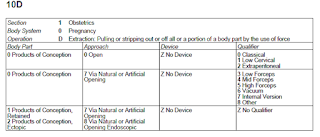
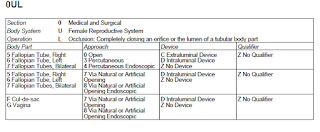
In regard to the “accepted” clinical standard, ACOG published the ACOG committee opinion #620 in January 2015,  in their eyes, ovarian cancer may originate in the fallopian tube leading to a new standard of care,  At this time they are pursuing more research, but agreed that if a patient was in for a tubal ligation, to excize the entire tube rather than only a portion of the tube for elective sterilization.  This is now considered a tubal ligation via salpingectomy.

Unfortunately, for coders; there is no CPT code for this idea of reporting a prophylactic salpingectomy at athe time of tubal ligation.  CPT gives us a code for “salpingectomy”  or “tubal ligation”   ACOG has given the physicians/surgeons coding options for this type or clinical care and reporting.

In querying ACOG as to how should reporting/coding be done, they have stated that salpingectomy code 58700 should NEVER be used to report a sterilization procedure of any sort.  The rationale behind this is that the RVU values of the salpingectomy code include the pathological changes and additional risk included in those changes that cause complications such as blocked tubes, adhesions, or even benign or neoplastic effects.   Their recommendation for coding is you code as per the “intent” of doing a tubal ligation.  In the request of clarity and transparency in coding and documentation, on the “notes” line of your claim form you should include a brief statement “tubal ligation via salpingectomy”.


Issue:  Facility codes as “Salpingectomy”; Physician codes as “Tubal Ligation”

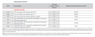
The issue has come up that when hospital inpatient coders code for a “tubal ligation via salpingectomy”  we are seeing these coded via the encoder as a standard salpingectomy – and the diagnosis appended to the facility claim is till sterilization.    This is problematic as you can see below”
 
1)      The physician’s billing and the facility billing do not match (cpt codes)

2)      The facility has been reimbursed at a higher RVU value rate for a salpingectomy than what is reimbursed for a traditional tubal ligation

3)      The physician and/or facility may be denied payment by a third party payer (insurance carrier) due to the claims do not ‘match’. 

4)      The patient ends up with a large “bill” from the non-payment of either the physician billing or the hospital/facility billing due to this mis-match on the claims.
 
5)      The facility claims they have coded correctly – as they do not recognize ACOG specialty guidelines for billing of facility charges for OB and GYN services. 


… And we have very complicated issues….

When this issue came up, the physician office was informed by the facility that they had “coded the procedure” incorrectly, and to immediately file a corrected claim showing “salpingectomy”  instead of “Tubal ligation”  as the facility had not been paid. 

The physician office countered this inquiry back to the facility; stating they were following the coding direction of ACOG and the clinical rationale behind performing salpingectomy vs/tubal ligation, and also the reimbursement rationale of why the physician should not be entitled to a higher reimbursement RVU value for this service.   

As you can see below, the RV values are significantly higher for the traditional salpingectomy than for the tubal ligation(s).  Also, the edits, show the salpingectomy is subject to 10 post op/ follow up days, and the tubal ligation(s) are subject to 90 post op follow up days.  This will also impact how care is delivered to the patient post surgery.


(Salpingectomy)
(Tubal Ligations)


The facility coder then stated that when they ran the scenario through the encoder, it directed them to code the salpingectomy.  Unfortunately, the encoder does not take into consideration the “reason” for the salpingectomy was for contraception (elective) not medically indicated (eg disease of the tube).  In addition the facility stated that CPT, also does not have a variance, or guideline for coding the “tubal ligation” code when a “salpingectomy” is performed.

Both sides of this issue have valid reasons for coding the way they did.  Unfortunately, the facility has not been paid, and has turned this billing over to patient responsibility.  Both side of this issue feel that the coding was coded correctly.  However, all coders should also adhere to the AHIMA and/or AAPC standards of ethical conduct when coding.   

AHIMA Standards of Ethical Coding are: (AHIMA link)
1.      Apply accurate, complete, and consistent coding practices for the production of high-quality healthcare data.

2.      Report all healthcare data elements (e.g. diagnosis and procedure codes, present on admission indicator, discharge status) required for external reporting purposes (e.g. reimbursement and other administrative uses, population health, quality and patient safety measurement, and research) completely and accurately, in accordance with regulatory and documentation standards and requirements and applicable official coding conventions, rules, and guidelines.

3.      Assign and report only the codes and data that are clearly and consistently supported by health record documentation in accordance with applicable code set and abstraction conventions, rules, and guidelines.

4.      Query provider (physician or other qualified healthcare practitioner) for clarification and additional documentation prior to code assignment when there is conflicting, incomplete, or ambiguous information in the health record regarding a significant reportable condition or procedure or other reportable data element dependent on health record documentation (e.g. present on admission indicator).

5.      Refuse to change reported codes or the narratives of codes so that meanings are misrepresented.

6.      Refuse to participate in or support coding or documentation practices intended to inappropriately increase payment, qualify for insurance policy coverage, or skew data by means that do not comply with federal and state statutes, regulations and official rules and guidelines.

7.      Facilitate interdisciplinary collaboration in situations supporting proper coding practices.

8.      Advance coding knowledge and practice through continuing education.

9.      Refuse to participate in or conceal unethical coding or abstraction practices or procedures.

10.  Protect the confidentiality of the health record at all times and refuse to access protected health information not required for coding-related activities ( examples of coding-related activities include completion of code assignment, other health record data abstraction, coding audits, and educational purposes).

11.  Demonstrate behavior that reflects integrity, shows a commitment to ethical and legal coding practices, and fosters trust in professional activities.

AAPC Code of Ethics (Link to AAPC ethics documents)

It shall be the responsibility of every AAPC member, as a condition of continued membership, to conduct themselves in all professional activities in a manner consistent with ALL of the following ethical principles of professional conduct:
      • Integrity
      • Respect
      • Commitment
      • Competence
      • Fairness
      • Responsibility


Adherence to these ethical standards assists in assuring public confidence in the integrity and professionalism of AAPC members. Failure to conform professional conduct to these ethical standards, as determined by AAPC's Ethics Committee, may result in the loss of membership with AAPC.

AAPC and its Ethics Committee does not function as a judiciary body. Any complaint to AAPC should have a foundation in law (for example, someone has been found guilty of fraud or has been placed on the Medicare Exclusion List) or a foundation in AAPC administrative rules (for example, counterfeit CEUs or a member using credentials falsely


Resolution?

The resolution for this case was for the facility to send an appeal back to the 3rd party carrier showing the rationale for each side of the issue.  (Eg; why the physician/surgeon/provider coded as a “tubal ligation via salpingectomy”  and the facility rationale for how the “computerized encoder”  directed the facility to code as a traditional salpingectomy)

At this time, the burden of payment now lies squarely on the 3rd party insurance payer.  While this is not a perfect solution, it clearly shows that a resolution can be had, but both the physician provider and the facility have a vital stake in needing reimbursement, yet have completely different ideas as to what is “right” and “wrong” with how this should be coded.

In the ever-changing world of medicine, these situations are becoming more and more common.  New tools are being developed for usage in operative procedures, new clinical information is now being discovered via scientific medical research.  CPT procedures, ICD10 pcs,  and encoders all struggle with maintaining the database and guidelines for each, based upon these clinical and scientific developments for appropriate patient care.

In regard to the contraceptive vs/medically indicated issue; many third party payers do not pay for contraceptive procedures, but they do pay for medically necessary procedures (such as a salpingectomy)  when there is a diagnostic reason.   In these cases, it is best to contact the 3rdparty payer/insurancecarrier in advance to verify what coverage the  patient may have.  . If coverage is not available or uncertain, be prepared to have the patient sign an advanced beneficiary notice and/or request payment in full at the time services are rendered.   

It is also advisable to contact and communicate with the facility to inform them of your “anticipated” coding prior to a surgery/procedure being performed.   All parties should be informed of the intent and “pre-authorize/pre-code” for the procedure.  These should include
a)      Physician Office, coder, biller, and physician
b)      Facility/Hospital coder, and encoder manager
c)      Patient
d)      Third party insurance payer/pre-authorization

In conclusion, the above scenario is not the only surgical scenario that this may happen with.  This can happen when a physician may perform what they deem as “medically necessary” and the facility deem as “cosmetic”.. so communication becomes a critical issue in these cases.  Resolution for these types of issues take perseverance and the willingness to understand the coding rationale on both sides to come to a good overall solution for all parties involved.


Editor’s note: Lori-Lynne A. Webb, CPC, CCS-P, CCP, CHDA, CDIP, COBGC and ICD10 cm/pcs Ambassador/trainer is an E&M, and Procedure based Coding, Compliance, Data Charge entry and HIPAA Privacy specialist, with over 20 years of experience.  Lori-Lynne’s coding specialty is OB/GYN office & Hospitalist Services, Maternal Fetal Medicine, OB/GYN Oncology, Urology, and general surgical coding.  She can be reached via e-mail at webbservices.lori@gmail.com or you can also find current coding information on her blog site: http://lori-lynnescodingcoachblog.blogspot.com/.  


Zika Virus - A Q&A Primer - Info on Zika is changing quickly - here's what I know as of today (03/02/2016)

$
0
0
This is the most current article that I wrote for Justcoding.com.  It is also free to access on their website.  However, I suggest becoming a full-subscription member, as they have a huge amount of resources and information available.  :) 


********************************************************************************************************

Zika Virus -  A Q&A Primer
by Lori-Lynne A. Webb, CPC, CCS-P, CCP, CHDA, COBGC, CDIP


What is Zika?

According to the Center for Disease Control (CDC)  this is the officialdefinition:

The Zika virus is a mosquito-transmitted infection related to dengue, yellow fever and West Nile virus. It was discovered in the Zika forest in Uganda in 1947 and is common in Africa and Asia.  It did not begin spreading widely in the Western Hemisphere until last May, when an outbreak occurred in Brazil.


A bit of clinical background

This is information direct from the American Congress of Obstetricians and Gynecologists (ACOG)  and the Society of Maternal and Fetal Medicine  (SMFM)

The virus spreads to humans primarily through infected Aedes aegyti mosquitoes. Once a person is infected, the incubation period for the virus is approximately 3-12 days. Symptoms of the disease are non-specific but may include fever, rash, arthralgias, and conjunctivitis. It appears that only about 1 in 5 infected individuals will exhibit these symptoms and most of these will have mild symptoms. It is not known if pregnant women are at greater risk of infection than non-pregnant individuals.

Zika during pregnancy has been associated with birth defects, specifically significant microcephaly. Transmission of Zika to the fetus has been documented in all trimesters; Zika virus RNA has been detected in fetal tissue from early missed abortions, amniotic fluid, term neonates and the placenta. However, much is not yet known about Zika virus in pregnancy. Uncertainties include the incidence of Zika virus infection among pregnant women in areas of Zika virus transmission, the rate of vertical transmission and the rate with which infected fetuses manifest complications such as microcephaly or demise. The absence of this important information makes management and decision making in the setting of potential Zika virus exposure (i.e. travel to endemic areas) or maternal infection, difficult. Currently, there is no vaccine or treatment for this infection.

The ACOG and SMFM put forth guidelines for testing of pregnant women, and the laboratory tests are being done exclusively though the guidance of the CDC at the level of the local and state health departments.  Many states in the US are developing guidelines to help in identifying who has been exposed, and where an outbreak may take place. 

Currently the testing being done is a “Zika” serology IgM testing assay.  The reports have been being reported out as “likely positive”, “Inconclusive” and “likely negative”  .  Unfortunately, the labs do not know and gannot guarantee the sensitivity of the IgM assay.


Symptoms of Zika

 Below is a listing of all the known symptoms of Zika virus as put forth by the CDC, however, there may be more that are noted as the Zika Virus becomes more studied in all individuals. Zika is still a virus, and not a bacterial infection, and currently there is not vaccine to prevent it, or a specific medication or antibiotic to treat it with. 

• About 1 in 5 people infected with Zika virus become ill (i.e., develop Zika).

• The most common symptoms of Zika are fever, rash, joint pain, or conjunctivitis (red eyes). Other common symptoms include muscle pain and headache. The incubation period (the time from exposure to symptoms) for Zika virus disease is not known, but is likely to be a few days to a week.

• The illness is usually mild with symptoms lasting for several days to a week.

• People usually don’t get sick enough to go to the hospital, and they very rarely die of Zika.

• Zika virus usually remains in the blood of an infected person for about a week but it can be found longer in some people.


Risks of Zika in Pregnant Women and in their sexual  partners

Normally Zika virus is transmitted through a mosquito bite, however, the Zika virus can be transmitted from a pregnant mother to her unborn fetus during the time of pregnancy and possibly around the time of birth.  It has been noted that Zika virus has been noted in all trimesters of pregnant women, and may possibly be transmitted during the birth process.  Sexual transmission of the Zika virus can also occur, however there is limited data, but the CDC has stated that if the patient fears they are infected with the Zika virus to reduce the risk of sexual transmission via abstinence and/or usage of condoms.

Women are not the only ones at risk of contracting Zika virus.  Men who have traveled to an area of active Zika virus, or who live in these areas may become infected with the Zika virus too.  The CDC has not completely determined if the Zika virus can be transmitted sexually, so the recommendation for men is if you are symptomatic or have a confirmed case of Zika virus, condoms or abstinence is still a best practice.  However, it remains uncertain if the mirus persisits in semen even if no longer  detectible in the blood.


Fetal Evaluation for possible exposure to Zika

Ultrasound exami is the primary recommendation for pregnant mothers who have been exposed to zika virus.  The Ultrasound examinations should focus on development of the fetal brain with intracranial calcifications and microcephaly.  Micocephay has been the most frequently reported adverse fetal complication  in women who have had the virus while pregnant

SMFM is recommending not only blood tests for pregnant women who have been exposed, but also consider performing serial ultrasound, as frequently as every 3-4 weeks.   By obtaining the additional ultrasounds, this would be considered ongoing surveillance.  Considering the history of Zika virus and complications to the fetus  due to this infection is not known.  In addition,  the time from exposure and infection from Zika  to  exhibiting full-blown clinical manifestations is unknown.

The CDC, ACOG and SMFM have put out a number of clinical flow algorhythms for usage with patients’ that have been exposed or live in an area where Zika as been prevalent.  However, this is so new, that these recommendations may change very quickly.   


Case Study and Coding Consideration


Case #1:
An asymptomatic pregnant woman at 19 weeks gestation, presents to her OB office for her regularly scheduled OB prenatal visit.  She informs the receptionist of the possibility she has been exposed to Zika. She has a history of travel to Mexico between 16+0 and 16+5-weeks. She has noted mosquito bites over both legs (calf area).  The bites do not appear infected, and look as if they are resolving.  Patient states they no longer itch, and does not report any other complaints but her ongoing pregnancy related fatigue.  The physician performs a comprehensive history, a comprehensive exam, and will have labs drawn for Zika to be sent to the local district health office.  In addition, the physician decides to perform a baseline screening ultrasound exam to follow up from the patient’s first trimester ultrasound anatomy exam from 1 month ago. 

Coding Consideration: 
CPT: 
99214-25 E&M  - 
76816 Ultrasound 
36415 Venipuncture/Lab Draw

ICD-10: 

O26.812   Pregnancy related exhaustion and fatigue (2ndtrimester)
Z20.828    Contact with and (suspected) exposure to other viral communicable        diseases (Zika Virus)
S80.861A  Insect bite of rt lower leg initial encounter
S80.862A  Insect bite of lt lower leg initial encounter
Z3A.19      19 weeks gestation of pregnancy

Rationale:  The  E&M visit would be coded, as it is separately identifiable  “outside” the normal pregnancy antenatal care.  (A Zika virus exposure is not considered “normal obstetric care”)  the follow-up ultrasound/baseline ultrasound is coded for comparison to the previously performed 1st trimester ultrasound.  The venipuncture is the only thing chargeable, as the blood was drawn, and sent out to the health district for testing.  The sequencing of the pregnancy diagnosis is primary based upon the ICD-10 pregnancy guidelines.


ACOG’s Quick Zika Q&A

Q1.  True or False. Pregnant women are at greater risk of infection with the Zika virus than nonpregnant women.
A:   False - According to a practice advisory from ACOG and SMFM, “It is not known if pregnant women are at greater risk of infection than non-pregnant individuals.”


Q2.  Once a person is infected with the Zika virus, what is the approximate incubation period for the virus?
A:.   3 to 12 days - Following infection with the Zika virus, the incubation period is approximately 3 to 12 days


Q3.  The Zika virus spreads to humans primarily through infected Aedes aegypti mosquitoes. Which of the following symptoms may be associated with the virus?
Fever
Rash
Arthralgia
Conjunctivitis
All of the above       

A.   Although symptoms associated with the Zika virus are non-specific, they may include fever, rash, arthralgia, and conjunctivitis. (eg all of the above)

Q4. In which trimester(s) has transmission of Zika been documented?

A. All trimesters -- The transmission of the Zika virus has been documented in all trimesters


Wrap up

At this time, there are still a number of unanswered questions in regard to the Zika virus.  However, there is no vaccine currently available, so it is recommended that precaution be taken to avoid exposure to mosquito bites from areas where the Zika virus is prevalent.  In the United States and worldwide expert epidemiologists are helping to set forth useful clinical guidelines for identifying and managing patients who have been exposed and currently have the Zika virus.  At this time, clinical guidelines are calling for blood tests to be run, and screening ultrasound should be performed on pregnant patients to screen for possible fetal anomalies related to fetal brain development in infected female patients.

When coding, carefully review to see if the physician or provider is stating whether the patient truly has the Zika virus as a diagnosis, or if they are only “screening” for the Zika virus in light of an exposure to the virus. (either through mosquito bite, or sexual transmission).  

In addition, currently, ICD-10 does not have a specific code to identify Zika virus. Usage of code B33.8 Other specified viral diseases, would be appropriate.  However, If the patient is diagnosed with the Zika virus and has fever with it, then it may be appropriate to use code A92.8 – Other specified mosquito-borne viral fevers.   If the patient is pregnant, then usage of ICD-10 code 098.5X “other viral diseases complicating pregnancy, childbirth and the puerperium,” (be sure to use the most specific trimester as the additional character) would be the most appropriate. 

If in doubt about the clinical documentation, be sure to query the provider to obtain clarity on the diagnosis noted in the medical record. 


References:
www.acog.org/
www.cdc.gov/zika


Editor’s note: Lori-Lynne A. Webb, CPC, CCS-P, CCP, CHDA, CDIP, COBGC and ICD10 cm/pcs Ambassador/trainer is an E&M, and Procedure based Coding, Compliance, Data Charge entry and HIPAA Privacy specialist, with over 20 years of experience.  Lori-Lynne’s coding specialty is OB/GYN office & Hospitalist Services, Maternal Fetal Medicine, OB/GYN Oncology, Urology, and general surgical coding.  She can be reached via e-mail at webbservices.lori@gmail.com or you can also find current coding information on her blog site: http://lori-lynnescodingcoachblog.blogspot.com/.  


Understanding Bariatric Surgery: CPT and Surgical Interventions

$
0
0

June 19, 2016
 Originally from my HCPro article

In our society, and medical community, the disease of obesity is considered a major health problem. Unfortunately, the disease process of obesity continues to be a major risk factor for the diagnoses in  many other diseases such as diabetes, hypertension, sleep apnea, arthritis, and many, many more.  Obesity is also medically associated with significant morbidity and mortality risk factors when any type of surgical or operative intervention, or even non-surgical hospitalization is necessary.
Most medical providers define and document obesity by the measurement of body mass index (BMI). The BMI is calculated by dividing a patient's mass (kg) by his or her height (m2). A normal BMI is considered in the range of 18.5-24.9 kg/m2. A BMI of 25-29.9 kg/m2 is considered overweight. A BMI of 30 kg/m2 or greater is classified as obese; this classification is further subdivided into class I, II, or III obesity.  In ICD-10cm, obesity and BMI are now easily identifiable, and should be documented in the patients’ records when obesity is being treated as a stand-alone diagnosis, or as part of a diagnosis with other disease processes that are impacted by obesity.   The ICD-10 codes Z68.xx should be coded in addition to the diagnosis of obesity in the medical record and on your insurance claims. 
Bariatric Surgery Origins
The first effective surgery for obesity in the United States was performed in 1954.  This controversial surgery introduced the jejunoileal bypass.  This “weight loss” surgery was met with controversy, as it did have a large amount of complications, such as extreme malnutrition.   In addition to malnutrition, patients also developed  serious complications secondary to the malabsorption (eg diarrhea, vomiting, eg)  and many required reversal of the bariatric procedure.  These initial complications in the infancy of bariatric medicine, provided the impetus for physicians and surgeons to search for better surgical interventions.  As surgical procedures have progressed and become surgically safer, and with less complications, there has been more acceptance from medical physicians who care for obese patients.  These providers are able to provide better education to the patient,  if a surgical intervention is warranted for morbid obesity diagnoses .   In addition, with better bariatric surgical procedures, especially those that are less invasive,  patients ultimately  have the opportunity for surgical success of elimination of an obesity diagnosis.
Currently, there are four basic concepts/options of choices for patients and physicians to decide upon when moving forward with bariatric surgery:
·         Gastric restriction with adjustable gastric banding  (eg, sleeve gastrectomy)
  • Sleeve gastrectomy
    • In a sleeve gastrectomy, part of the stomach is separated and removed from the body. The remaining section of the stomach is formed into a tube like structure. This smaller stomach cannot hold as much food. It also produces less of the appetite-regulating hormone ghrelin, which may lessen your desire to eat. However, sleeve gastrectomy does not affect the absorption of calories and nutrients in the intestines.

  • Gastric restriction with mild nutritional malabsorption (eg Roux-en-Y gastric bypass)
    • The Roux-en-Y gastric bypass,
      • A  small stomach pouch is created with a stapler device and connected to the distal small intestine. The upper part of the small intestine is then reattached in a Y-shaped configuration.

  •  “Combination” surgery, that includes both mild gastric restriction and malabsorption (duodenal switch)
    • Sleeve gastrectomy with duodenal switch
      • In this procedure, the physician performs a “sleeve gastrectomy” which includes a duodenal switch.
      • The stomach is resected and "tubulized" with a residual volume of about 150 ml. This gastric reduction is the food intake restriction component.  The stomach itself, is then resected from the duodenum and connected to the distal part of the small intestine.  Once that is completed, the duodenum and the upper part of the small intestine are reattached to the rest at about 75–100 cm from the colon.

·         Laparoscopic adjustable gastric banding
·         “Lap Band” surgery
The laparoscopic adjustable gastric banding procedure, also known as the “Lap Band” surgery,  uses a laparoscopic approach to insert a band containing an inflatable balloon to be placed around the upper part of the stomach then fixed in place. This procedure allows a small stomach pouch to be “created”  above the band with a very narrow opening to the rest of the stomach.

·         A port is then placed under the skin of the abdomen. A tube connects the port to the band. Once in place, the surgeon or physician can adjust the band itself by injecting or removing fluid through the port.  This allows, the balloon to be inflated or deflated to adjust the size of the band, therefore restricting the amount of food that the stomach can hold.  This allows the patient to feel full sooner, but it doesn't reduce the absorption of calories and nutrients.


As with any of the above generalized components of bariatric surgery, there are many variations to each of the above four main types of surgical intervention.   CPT has done a terrific job of giving coders a wide selection of CPT codes to choose from to describe these surgical interventions.   In addition to the CPT codes, the surgeons have also abbreviated the surgeries as below in this table that the  American Society for Metabolic and Bariatric Surgery (ASMBS) put together as a helpful guide for coders to use.


Open Procedures
VBG
Gastric restrictive procedure, without gastric bypass, for morbid obesity; vertical-banded gastroplasty
43842
AGB
Gastric restrictive procedure, without gastric bypass, for morbid obesity; other than vertical-banded gastroplasty
43843
BPD/DS
Gastric restrictive procedure, with partial gastrectomy, pylorus-preserving duodenoileostomy (50 to 100 cm common channel) to limit absorption (BPD/DS)
43845
RYGB (proximal)
Gastric restrictive procedure, with gastric bypass for morbid obesity; with short limb (less than 150 cm) Roux-en-Y gastroenterostomy
43846
RYGB (distal)
Gastric restrictive procedure, with gastric bypass for morbid obesity; with small intestine reconstruction to limit absorption
43847
Revision RYGB
Revision, open, of gastric restrictive procedure for morbid obesity, other than adjustable gastric restrictive device (separate procedure)
43848
BPD
Gastrectomy, partial, distal; with Roux-en-Y reconstruction
43633

Laparoscopic Bypass Procedures

RYGB (proximal)
Laparoscopy, surgical, gastric restrictive procedure; with gastric bypass and Roux-en Y gastroenterostomy (Roux limb 150 cm or less)

43644
RYGB (distal)
Laparoscopy, surgical, gastric restrictive procedure; with gastric bypass and small intestine reconstruction to limit absorption

43645
Lap DS, Lap revisions
Lap sleeve gastrectomy
Unlisted laparoscopy, stomach
43659

Laparoscopic Gastric Restrictive Procedures

Lap adjustable gastric band and port implantation

Implantation of adjustable gastric band and port, [Laparoscopic]
43770
Lap Sleeve Gastrectomy
Laparoscopy, surgical, gastric restrictive procedure; longitudinal gastrectomy (i.e., sleeve gastrectomy)
43775


Let’s take a look at the operative reports

The first operative report is of a traditional laparoscopic sleeve gastrectomy used by CPT code 43775 -  then we have another laparoscopic sleeve gastrectomy that utilized a “robotic” assisted laparoscopic system for the same sleeve gastrectomy.  When coding for these be aware of what “tools” your provider is using if the procedure is being performed as a traditional laparoscopic surgery, or if the physician is utilizing a laparoscopic robotic system.

When coding these, the traditional operation will only require CPT code 43775; however, it you are utilizing a robotic system you should cod the 43775 as your first line item, then add HCPCS code S2900 at $0.00 to provide transparency to the codes and inform your insurance payers that the surgery was performed with a robotic laparoscope system.  Be aware that inclusion of the HCPCS code S2900 should not be billed as a stand-alone code, nor is it reimbursable for any extra revenue.  It is simply an “informational” code for the payers.  


Operative Report #1: Laparoscopic sleeve gastrectomy (traditional) 
Operative Report #2: DaVinci MIS (robotic)  laparoscopic sleeve gastrectomy
Operative Report #3: Laparoscopic (Lap-Band) gastric band placement
Operative Report #4: Laparoscopic removal of LAP-BAND, due to pregnancy (enlarged uterus)


As you review these operative reports, you will notice that these are all laparoscopic.  At this time, laparoscopic adjustable gastric banding is considered the least invasive surgical option for morbid obesity.  In addition, the laparoscopic sleeve gastrectomy which is also considered a viable surgical option, is also less invasive than a traditional open procedure with a quicker recovery time.  The Lap Band procedure is potentially reversible.  The laparoscopic sleeve gastrectomy is non-reversable. 

ICD-10 and Bariatric Surgery Status

The ICD-10-CM code Z98.84 Bariatric Surgery Status refers to the presence of any of these type of synonyms used in the clinical documentation of the medical record. 
·         bariatric surgery status 
·         gastric banding status  gastric bypass status for obesity
·         obesity surgery status
  • History of bariatric (weight loss) surgery
  • History of bariatric surgery
  • History of diabetes mellitus resolved post bariatric surgery
  • History of diabetes mellitus resolved post bariatric surgery (situation)
  • History of diabetes mellitus resolved post gastric bypass
  • History of diabetes mellitus resolved post gastric bypass (situation)
  • History of gastric bypass
  • Presence of laparoscopic band/ or presence of laparoscopic gastric banding device
If the patient is pregnant, and the patients’ bariatric surgery status is affecting the pregnancy, the ICD-10-CM refers us to use these codes as outlined below.  However, the physician should be sure to notate that the bariatric surgery status is complicating the pregnancy, and in what matter the complications exist.  The provider should clearly reflect any complications to the pregnancy related to the bariatric surgery status. 
O99.84 Bariatric surgery status complicating pregnancy, childbirth and the puerperium
·         O99.840 Bariatric surgery status complicating pregnancy, unspecified trimester
·         O99.841 Bariatric surgery status complicating pregnancy, first trimester
·         O99.842 Bariatric surgery status complicating pregnancy, second trimester
·         O99.843 Bariatric surgery status complicating pregnancy, third trimester
·         O99.844 Bariatric surgery status complicating childbirth
·         O99.845 Bariatric surgery status complicating the puerperium

As a coder, good documentation from your providers help ensure you are able to clearly code and report the operative session(s), with the diagnosis of obesity and all additional diagnoses that are impacted by the obesity (medical necessity).  All of these criteria go hand in hand with good quality patient care and correct coding and billing of claims. By working closely with your providers, you can ensure good clean claims, and reduce your overall risk of audit inquiry and financial recoupment of paid claim services.

Lori-Lynne A. Webb, CPC, CCS-P, CCP, CHDA, COBGC and ICD10 cm/pcs Ambassador/trainer is an E&M, and Procedure based Coding, Compliance, Data Charge entry and HIPAA Privacy specialist, with over 20 years of experience.  Lori-Lynne’s coding specialty is OB/GYN office & Hospitalist Services, Maternal Fetal Medicine, OB/GYN Oncology, Urology, and general surgical coding.  She can be reached via e-mail at webbservices.lori@gmail.com or you can also find current coding information on her blog site: http://lori-lynnescodingcoachblog.blogspot.com/.  
*********************************************************************************************

Operative Report #1
Laparoscopic sleeve gastrectomy (traditional) 
Patient is prepped and all antiembolic precauations are undertaken and appropriate preop antibiotics are administered via IV. A 12-mm optical trocar is placed under direct vision approximately 15 cm below the xiphoid and 3 cm to the left of midline
A 45-degree angled laparoscope is placed through the port into the peritoneal cavity and 12-mm port is placed in the left lateral flank, medial to the edge of the colon with the patient in a supine position and at the same level as the periumbilical port. Next, a 5-mm trocar port is placed along the left subcostal margin between the xiphoid process and the left flank port. Another 12-mm port is placed in the right epigastric region and a fourth 12 mm port was placed in the mid-epigastric region caudal and medial to the previous port. The liver is elevated and this provides adequate visualization of the entire stomach .
The pylorus of the stomach is then identified and the greater curve of the stomach elevated. An ultrasonic scalpel is then used to enter the greater sac via division of the greater omentum. The greater curvature of the stomach is then dissected free from the omentum and the short gastric blood vessels using the laparoscopic ultrasonic scalpel.
The dissection is started 5 cm from the pylorus and proceeds to the Angle of His .  A 9.8 mm gastroscope is then passed under direct vision through the esophagus, stomach, and into the first portion of the duodenum. The gastroscope is aligned along the lesser curvature of the stomach and used as a template to perform the vertical sleeve gastrectomy beginning 2 cm proximal to the pylorus and extending to the Angle of His.
An endoscopic linear cutting stapler is used to serially staple and transect the stomach staying just to the left and lateral to the endoscope. The gastrectomy is visualized with the endoscope during the procedure. The transected stomach, which includes the greater curvature, is completely freed and removed from the peritoneum through the left flank port incision . The staple line along the remaining tubularized stomach is then tested for any leak through insufflations with the gastroscope while the remnant stomach is submerged under irrigation fluid. The staple line is concurrently evaluated for bleeding both intraperitoneally with the laparoscope as well as intraluminally with the gastroscope. A 19-French Blake drain is left in the left upper quadrant along the sleeve gastrectomy staple line. Closure of the fascia t the left flank port site is performed with an absorbable suture on a transabdominal suture passer, to prevent bowel herniation.  We did not close the fascial defects at the remaining port sites.
Patient is taken to PACU in good condition.

CPT code:
43775:  Longitudinal gastrectomy (ie sleeve gastrectomy)


Operative Report #2 
DaVinci MIS (robotic)  laparoscopic sleeve gastrectomy
The Veress needle technique was used to establish the pneumoperitoneum into the left hypochondrium. A 12 mm port was inserted 120 mm inferior and slightly left to the sternum for camera access. For the latter port, we used an extra large 150 mm long trocar The right 12 mm working port was positioned 6 cm from the midline trocar. The left 12 mm working port was located 6 cm to the left of the midline trocar. An 11 mm trocar was placed laterally to the left hypochondrium and an 8 mm da Vinci trocar was placed under the right hip as laterally as possible to allow liver retraction. The 8 mm da Vinci trocars were inserted through standard, disposable 12 mm trocars. This double-cannulation technique was used asstandard 12 mm trocars are required during the insertion of the staples. All trocars are inserted under direct visualization with the da Vinci system camera
We began recording the docking time of the Robot.  The robotic camera was locked last but was used to insert all robotic cannulas and instruments. The robotic cart was positioned over the patient’s head. Once the general setup was ready, the procedure began with myself using a grasper in the left hand and a modified harmonic scalpel in the right hand. The third da Vinci arm used another forceps in order to retract the liver from the 8 mm trocar placed in the right-hand side of the patient. The greater curvature of the stomach was sectioned at the lowest point in order to reach the lesser epiploic sac. During this stage of the procedure, we are completely robotic.   The division of the gastrocolic and gastrosplenic ligament continued exactly as in a standard LSG. With care, we ensure precision in the upper part of the stomach, and avoided any injury to the spleen and had adequate visualization of the vessels. Dissection continued to 5 cm from the pylorus following dissection of the upper part of the stomach.
Next, the assistant surgeon inserted a 32 Fr bougie to calibrate the sleeve. The anesthesiologist did not encounter any difficulty placing the bougie with the robotic bedside cart. A Echelon 60 Endopath stapler, endoscopic linear cutter straight, loaded with a green cartridge, was used to divide the stomach from the lowest tip of the greater gastric curvature;  5 cm proximally to the pylorus, towards the lateral edge of the bougie. This maneuver was performed twice. The right arm was again docked and the left robotic arm was switched to the left lateral 11 mm trocar. This maneuver allowed the decannulation of the right arm from the 12 mm trocar without moving the robot.   We then inserted a stapler loaded with blue cartridges to divide the sleeve up to the end of the upper part. The stomach was then removed from the cavity through the 12 mm trocar. A robotic continuous polypropylene suture (3/0) was used to oversew the entire sleeve staple line.. The first assist then filled the sleeve with diluted methylene blue to detect any leakage from the staple line.  No leaks were encountered, and operative session was complete.  Patient taken to PACU in good condition. 
CPT code:
43775:  Longitudinal gastrectomy (ie sleeve gastrectomy)
S2900: Surgical Techniques Requiring Use Of Robotic Surgical System (List Separately In Addition To Code For Primary Procedure)


Operative Report #3
Laparoscopic (Lap-Band) gastric band placement
The procedure consisted of laparoscopic placement of a gastric band (Lap-Band System), creating a proximal 15-mL pouch at the cardia.
The patient was positioned in an elevated recumbent position. The video monitor was located beyond the patient’s right shoulder.  Pneumoperitoneum was created using a Palmer-Veress needle. The 10-mm optical trocar was inserted first, 10 cm below the xiphoid notch. Then, three 10-mm cannulas were placed under the rib margin.  The fourth cannula on the left had a larger diameter (18 mm) to allow the introduction of the band. All cannulas were then shifted to the left when preoperative (re-review) ultrasound revealed an enlarged left liver lobe (>15 cm high) in the patient. A 10-mm liver retractor was inserted through a paraxiphoid cannula and the left lobe was elevated to expose the cardiac area and the diaphragmatic crus.
Gastric dissection started at the angle of the cardia by division of the phrenogastric ligament. We proceeded with the lap band procedure with a pars flaccida approach on the right side.  Dissection on the left side was identical to that performed on the right. Over the lesser omentum, we opened the peritoneal sheet close to the edge of the right crus, then gradually created a retrogastric tunnel reaching the left crus and the phrenogastric ligament. Thus avoiding tthe use of a balloon.  The band was secured by an anterior gastrogastric valve using four nonabsorbable seromuscular stitches.   .  This covered the anterior part of the band completely. A methylene blue dye test was carried out with no leaks detected.  The subcutaneous port components were then placed and verified as per our pre-operative marking.   Patient was taken to PACU in good condition. 
CPT Code: 43770: Laparoscopy, surgical, gastric restrictive procedure; placement of adjustable gastric band (gastric band and subcutaneous port components)


Operative Report #4
Laparoscopic removal of LAP-BAND, due to pregnancy (enlarged uterus)

INDICATION FOR PROCEDURE:
This is a 27-year-old female who approximately 3 years ago had an adjustable gastric band placed laparoscopically.  She did well and lost over 100 pounds and subsequently became pregnant with twins.  At approximately 22 weeks' gestation, she started having nausea and vomiting and could not hold food down.  She had some morning sickness in the first trimester, which resulted in multiple bouts of nausea and vomiting, which may have been the etiology of initial slip of her band.  Slip of the band was confirmed during upper GI swallow.  She was referred by Dr.____, with the aforementioned findings requesting in consultation. 

In consultation, it was recommended the band could be put back in place and/or removed, and the patient requested removal of the band.

DESCRIPTION OF PROCEDURE: the abdomen was prepped and draped in the normal sterile fashion, a transverse 1 cm incision was made in the right upper quadrant approximately 1-inch medial to the anterior axillary line and 1 to 1-1/2 inches below the costal margin.  A 5 mm Optiview port was then advanced through the subcutaneous tissue, abdominal wall muscle, and immediately upon advancing through the abdominal wall muscle, encountered the uterine muscle, at which point the blunt trocar was removed.  A different angle tried and subsequently again the uterus encountered.  At this point, an additional incision approximately 2 inches lateral to the incision very near the costal margin was made, and a 5 mm port was able to be placed in the abdomen and insufflated.  Two small muscular lacerations on the right upper portion of the uterus were noted.  Under direct visualization, a 15 mm port was placed in the left upper quadrant directed towards the esophageal hiatus in the midclavicular line approximately 2 cm inferior to the costal margin.  In the epigastrium very near the xiphoid and just deviated to the left, an additional 5 mm port was placed, and a liver retractor was placed, retracting the left lobe of the liver anteriorly.  The patient was placed in reverse Trendelenburg, and a 5 mm port was placed through the original attempted site placement.  All instruments were used in the upper third of the abdomen as the lower two thirds of the abdomen were completely taken up by the very large uterus.  The gastric band tubing was identified, and it was elevated.  Scar tissue of omentum and adipose tissue were divided over this and taken down through the point of the buckle, which was opened.  The band was then adequately freed, the tubing cut, and the buckle opened completely by pulling the tubing through.  The wide part of the locking portion of the buckle, which was anterior, was then divided, which allowed the band to be removed without pressure or difficulty.  It was pulled out through the 15 mm port site in 3 pieces.  The remaining tubing will be pulled out with the subcutaneous port when this is dissected from its left lateral position. 

The ports were then removed under direct visualization, noting no bleeding at any of the port sites.  The liver retractor had been removed prior to moving the ports under direct vision without injury to intraabdominal contents.  The fascia in the 15 mm port site was closed with a figure-of-eight stitch of 0 Monocryl.  The skin directly in the old incision very close to the port was infiltrated with local anesthetic, and a 3 cm incision was made dissecting down and identifying the port.  The port capsule and suture was then dissected free of surrounding tissue and removed along with the port and the tubing.  The skin was then closed at this site with simple interrupted buried sutures of 4-0 Monocryl as was the remainder of the laparoscopic sites.  The skin and all incisions were sealed with Dermabond.

CPT code: 43774 Laparoscopy, surgical, gastric restrictive procedure; removal of adjustable
gastric restrictive device and subcutaneous port components


Tips for Coding and Documenting for Bariatric Surgery in ICD-10pcs - Inpatient

$
0
0


This is from my HCPro article  published June 2016
In last weeks HCPro outpatient article we addressed tips for coding in the physician office, and the challenges with that side of medicine that affects both the physician and the physician office or group practice.   In this article, we are addressing the inpatient side. 

As discussed in the outpatient article, the disease of obesity is considered a major health problem In the US.  Unfortunately, the disease process of obesity continues to be a major risk factor for the diagnoses in many other diseases such as diabetes, hypertension, sleep apnea, arthritis, and many, many more.  Obesity is also medically associated with significant morbidity and mortality risk factors when any type of surgical or operative intervention(s), or even non-surgical hospitalization is necessary.
Most medical providers define and document obesity by the measurement of body mass index (BMI). The BMI is calculated by dividing a patient's mass (kg) by his or her height (m2). A normal BMI is considered in the range of 18.5-24.9 kg/m2. A BMI of 25-29.9 kg/m2 is considered overweight. A BMI of 30 kg/m2 or greater is classified as obese; this classification is further subdivided into class I, II, or III obesity.  In ICD-10cm, obesity and BMI are now easily identifiable, and should be documented in the patients’ records when obesity is being treated as a stand-alone diagnosis, or as part of a diagnosis with other disease processes that are impacted by obesity.   The ICD-10 codes Z68.xx should be coded in addition to the diagnosis of obesity in the medical record and on your insurance claims

As we have been perfecting our ICD-10pcs coding skills with the ICD-10 tables;  Let’s take a quick look again at the basics of code construction. 

·         All ICD-10-PCS codes have seven digits, each digit representing a specific character associated with procedures.
·         Code assignment in ICD-10-PCS is a process of “constructing” the code by selecting values from the ICD-10 pcs code tables for each of the seven standard characters.
·         The first three characters identify the code table that is used to complete the remaining four characters.


The basics of bariatric ICD-10-pcs code selection
·         1: Section:  For bariatric procedures; the appropriate section is 0-Medical and Surgical.

·         2: Body System:  Bariatric procedures involve the stomach and intestines, so code tables need to be referenced from; D-Gastrointestinal System.

·         3: Root Operation:  When coding for the Root operation, in bariatric surgery, these are assigned according to the objective of the procedure.  There are standard definitions to be reviewed in ICD-10 for root operations.  When choosing the root operation, and the specific procedure that the physician is going to perform, there are three root operations that are most commonly used in bariatric coding.

1.       Bypass: Altering the root of passage for the contents of a tubular body part, eg, Roux-en-Y gastric bypass
2.       V-Restriction: Partially closing an orifice or the lumen of a tubular body part, eg, gastric banding
3.       B-Excision: Cutting out or off, without replacement, a portion of a body part, eg, sleeve gastrectomy

§  Note:  that because the procedure's objective is the defining factor in assigning the root operation, some procedures that are not associated with bariatric coding may also use the same ICD-10-PCS code.

§  Note: The physician is not expected to document using ICD-10-PCS code descriptions. It is your responsibility as a coder to determine what the physician's operative note documentation equates to in terms of ICD-10-PCS.  AHIMA has stated that coder is not required to query the physician in these circumstances.

·         4: Body Part:   In the respective ICD-10 pcs code tables the specific body part values that are available for you to choose from are for stomach, duodenum, and ileum.

·         5: Approach:  The approach used for the bariatric surgical procedures performed are:
o   Via laparotomy use 0-Open.
o   Via laparoscopy use 4-Percutaneous Endoscopic.

·         6: Device:  Interestingly in bariatric surgery, the device character is not used for surgical instruments that accomplish the procedure.  The device character is used to describe the devices that remain in the patient's body after the procedure is completed.  (eg, implanted devices) 
o   For a Gastric banding procedures, the coder will use
§  C-Extraluminal Device because the band encircles the lumen of the stomach from the outside.
o   If you are coding other bariatric procedures,
§   Z-No Device is most common choice when coding.

·         7: Qualifier: Qualifiers add further information to the ICD-10pcs code choice.
o   For therapeutic procedures, the most common qualifier is Z-No Qualifier.
o   For bypass procedures, the qualifier identifies the body part being bypassed to
§  eg…  re-routing the digestive tract from the stomach directly to the ileum you would use the  uses the qualifier B-Ileum.

Operative Report #1 

Laparoscopic (Lap-Band) gastric band placement
The procedure consisted of laparoscopic placement of a gastric band (Lap-Band System), creating a proximal 15-mL pouch at the cardia.
The patient was positioned in an elevated recumbent position. The video monitor was located beyond the patient’s right shoulder.  Pneumoperitoneum was created using a Palmer-Veress needle. The 10-mm optical trocar was inserted first, 10 cm below the xiphoid notch. Then, three 10-mm cannulas were placed under the rib margin.  The fourth cannula on the left had a larger diameter (18 mm) to allow the introduction of the band. All cannulas were then shifted to the left when preoperative (re-review) ultrasound revealed an enlarged left liver lobe (>15 cm high) in the patient. A 10-mm liver retractor was inserted through a paraxiphoid cannula and the left lobe was elevated to expose the cardiac area and the diaphragmatic crus.
Gastric dissection started at the angle of the cardia by division of the phrenogastric ligament. We proceeded with the lap band procedure with a pars flaccida approach on the right side.  Dissection on the left side was identical to that performed on the right. Over the lesser omentum, we opened the peritoneal sheet close to the edge of the right crus, then gradually created a retrogastric tunnel reaching the left crus and the phrenogastric ligament. Thus avoiding tthe use of a balloon.  The band was secured by an anterior gastrogastric valve using four nonabsorbable seromuscular stitches.  This covered the anterior part of the band completely. A methylene blue dye test was carried out with no leaks detected.  The subcutaneous port components were then placed and verified as per our pre-operative marking.   Patient was taken to PACU in good condition. 
Coding Choices:
ICD-10pcs code: 0DV64CZ
Previous ICD-9 Vol 3:  44.95
CPT code: CPT Code: 43770 Laparoscopy, surgical, gastric restrictive procedure; placement of adjustable gastric band (gastric band and subcutaneous port components


Now as we look at some of the coding options for an “open” and “laparoscopic’ bypass procedure you will note the same table is used, but they are 2 completely different codes as one is an “open procedure” the other is “laparoscopic”

• Gastric bypass from stomach to ileum, performed via laparotomy
0D160ZB Bypass stomach to ileum, open approach

• Gastric bypass from stomach to jejunum, performed via laparoscopy
0D164ZA Bypass stomach to jejunum, percutaneous endoscopic approach
*             


Diagnosis coding for bariatric medicine and bariatric surgery requires not only the definitions of the obesity, but notation of BMI.  Most often the obesity diagnosis will remain as the primary reason for bariatric surgery, but any co-morbidities will also play into the DRG that will affect the reimbursement for the facility where the bariatric surgery is being performed.

According to AHIMA, they suggest including this into your medical records for the clinical documentation when referencing obesity and bariatric surgery:

• Obesity
                - Morbid (severe)
° Due to excess calories
° With alveolar hypoventilation (Pickwickian syndrome)
 - Drug Induced
° Document drug
 - Other ° Due to excess calories, familial, endocrine

• Overweight
• Body Mass Index (BMI)
• Document any associated diagnoses/conditions
o   Hypertension
o   Type II Diabetes Mellitus
o   Dyslipidemia
o   Musculoskeletal, neurological or body size problems precluding or severely impairing quality of life (employment, family function or ambulation)
o   Life-threatening Cardiopulmonary Problems (sleep apnea, obesity-hypoventilations syndrome or obesity-related cardiomyopathy)
o   Coronary Artery Disease
o   Obesity-Related Cardiomyopathy
As we can see from this table below of 2016 “estimated” DRG’s and reimbursements for facilities, a bariatric surgery can be very lucrative for your facility.  The DRG assignments will need to be carefully reviewed when coding out bariatric surgery to obtain the highest appropriate DRG’s. 


Currently, there has been an increase in private insurance companies covering bariatric surgical procedures if the patient meets the standard criteria for morbid obesity.  However, some carriers may not cover it at all, and it may be a self-pay only option for the patient.  Medicare has been one of the primary payers that have approved bariatric surgery, with the resulting off-set of better health for the patient, and a reduced risk of long-term medical complications from the co-morbidities.

Medical necessity plays a huge part in a patient being able to undergo a bariatric surgery.  If the patient is morbidly obese and has a body mass index (BMI) of 40 or higher an insurance carrier is more likely to approve or pre-authorize a surgery.  Another criteria that may be imposed, is if the patient has been obese for the past five years or longer, and has
attempted, under a physician’s care;  other methods of weight loss for at least two years. These may include behavior modification, psychological evaluations, in addition to specifically proven medically regulated diets such as “Optifast”  “Medifast”  or even drug therapies such as orlistat (Xenical), lorcaserin (Belviq), phentermine and topiramate (Qsymia), buproprion and naltrexone (Contrave), and liraglutide (Saxenda).  If the patient has comorbidities such as hypertension, diabetes, sleep apnea, degenerative arthritis, and heart disease that increase the consideration of medical necessity for surgery.

In addition there are some patients in which they would not qualify for bariatric surgery. Absolute contraindications to bariatric surgery are active substance abuse and psychiatric personality disorders.  In addition, previous abdominal surgeries or previous bariatric procedures that were ineffective are not necessarily contraindications, but the patient may not be approved for more extensive bariatric surgery.  Some studies have borne out that procedures which alter the size of the stomach and restrict food intake, may exacerbate some eating disorder.  If the patient has a history of a true anorexia nervosa, they are generally considered not eligible for bariatric surgery.
As a coder, good documentation from your providers in the H&P  help ensure you are able to clearly code and report the operative session(s), with the diagnosis of obesity and all additional diagnoses that are impacted by the obesity (medical necessity).  All of these criteria go hand in hand with good quality patient care and correct coding and billing of claims. By working closely with your providers, you can ensure good clean claims, and reduce your overall risk of audit inquiry and financial recoupment of paid claim services.


Lori-Lynne A. Webb, CPC, CCS-P, CCP, CHDA, COBGC and ICD10 cm/pcs Ambassador/trainer is an E&M, and Procedure based Coding, Compliance, Data Charge entry and HIPAA Privacy specialist, with over 20 years of experience.  Lori-Lynne’s coding specialty is OB/GYN office & Hospitalist Services, Maternal Fetal Medicine, OB/GYN Oncology, Urology, and general surgical coding.  She can be reached via e-mail at webbservices.lori@gmail.com or you can also find current coding information on her blog site: http://lori-lynnescodingcoachblog.blogspot.com/.   

Diagnosis Coding for Obesity, BMI, when noted in the clinical record

$
0
0
Diagnosis Coding for Obesity, BMI, when noted in the clinical record
May 20, 2016

As a coder, we are faced with the challenges of reporting all diagnoses held within the medical record that the providers are currently addressing during an encounter with the patient.  The diagnosis of obesity is one of those difficult coding issues.  Obesity is a complicating factor in many areas of health care, and its effect upon care is multifold.    According to the National Institutes of Health (NIH), they define morbidobesity as:
·         Being 100 pounds or more above your ideal body weight.
·         Having a Body Mass Index (BMI) of 40 or greater.
·         Having a BMI of 35 or greater and one or more co-morbid condition.

High-risk comorbid conditions include the diagnoses of; Type 2 diabetes, life-threatening cardiopulmonary problems (egg, severe sleep apnea, Pickwickian syndrome, obesity-related cardiomyopathy), obesity-induced physical problems interfering with a normal lifestyle (e.g., joint disease treatable but for the obesity), and body size problems precluding or severely interfering with employment, family function, and ambulation.

In addition, mental status can also play a part in a patients’ obesity.  Mental status is a difficult diagnosis in and of itself, but can be another diagnosis that will need to be addressed if the physician notes the mental issues such as; severe depression, untreated or undertreated mental illnesses associated with psychoses, active substance abuse, bulimia nervosa, and socially disruptive personality disorders in addition to the obesity.   The Centers for Disease Control (CDC) states that over the last 30 years (as of 2009) that obesity is now considered to be “epidemic” in the United States and in adults 60 years and older is approximately 37% and 34% among women.  

The NIH breaks down obesity into “classes”
Class I Obesity = BMI 30.0 – 34.9 kg/m2
Class II Obesity = BMI 35.0 – 39.9 kg/m2
Class III Obesity = BMI ≥ 40 kg/m2

As a coder, by utilizing the information documented in the record, we can code the BMI from a dietitian's note, or from the physician’s documentation.  However, if the numeric BMI falls into the “class” status we can report and code this as a Class I, II, or III obesity state.  The obesity documentation still has to be clearly defined within the medical record.  With that, there should be a correlation from the physician to support the obesity code assignment, and how that is currently impacting the patients’ current care and ongoing plan.

The next coding challenge to coding of an obesity diagnosis is the notation of the word “morbid” obesity.   As we know from the NIH, the definition of such is defined, yet many physicians note in the record the words “patient is morbidly obese” but do not include any further information or documentation for the coder to adequately code the obesity diagnosis correctly for that particular patient.  A patient may not have all the criteria for being “morbidly obese” according to the NIH guideline, however, a physician may document that the patient is “morbidly obese” in the medical record.   If the documentation of an obesity diagnosis is a pertinent part of that patients’ care or reason for their medical encounter; the coder is obligated to record the diagnosis accurately and may need to query the provider and ask for clarification or additional information to clearly support the “morbidly obese” diagnosis.  In addition, Coding Clinic, fourth quarter 2005, stated that coders could code BMI based on notes from dietitians, but we should still be diligent in having this information corroborated by the physician in the record too. 

AHIMA has given us a quick tool to use when asking the physician to clarify a diagnosis related to obesity.  In the ICD10cm changes for codes; the listing below helps us give clarity to the physicians, to document what we need to have to clearly report an obesity diagnosis correctly.  In addition, a BMI only identifies the ratio of height to weight and there may be outside factors or other reasons that can alter a BMI “number, such as highly muscular people, pregnant or lactating women.  It is not appropriate to assume or make the correlation that someone is diagnostically obese from a high BMI nor considered diagnostically underweight from a low BMI.

        Obesity
Morbid (severe)
° Due to excess calories
° With alveolar hypoventilation (Pickwickian syndrome)
Drug Induced
° Document drug
Other
° Due to excess calories, familial, endocrine
        Overweight
        Body Mass Index (BMI)
        Document any associated diagnoses/conditions

From a coding perspective, documentation to support a diagnosis of overweight, obesity, and morbid obesity, obesity, should be clearly defined by the physician.  This documentation may include:

Ø  Diet discussed
Ø  Exercise encouraged
Ø  Gastric bypass surgery consult
Ø  Diet medication
Ø  Dietician referral and/or counseling
Ø  Weight loss program (i.e. gym membership)
Ø  Food log
Ø  Physiatrist referral

Obesity and Pregnancy

In April 2016, the American Congress of Obstetricians and Gynecologists (ACOG) defined what they consider obesity to be, and they closely follow the NIH guidelines.  ACOG defines the term “overweight” as having a body mass index (BMI) of 25–29.9.; and define the term “obesity” as having a BMI of 30 or greater.    ACOG has also noted that within the general category of obesity, there are three levels of “risk” go hand in hand with an increasing BMI:

        Lowest risk is a BMI of 30–34.9.
        Medium risk is a BMI of 35.0–39.9.
        Highest risk is a BMI of 40 or greater

ACOG has also confirmed that obesity during pregnancy puts the pregnant female at risk for several serious health problems such as:

        Gestational diabetes:
o   Gestational diabetes that is first diagnosed during pregnancy and can increase the risk of having a cesarean delivery.
o   Women who have had gestational diabetes also have a higher risk of having diabetes in the future, as do their children.
o   Obese women should be screened for gestational diabetes early in pregnancy and also may be screened later in pregnancy as well.

        Preeclampsia:
o   Preeclampsia is a high blood pressure disorder that can occur during pregnancy or after pregnancy.
o   It is a serious illness that affects a woman’s entire body.
o   The kidneys and liver may fail.
o   Preeclampsia can lead to seizures, a condition called eclampsia.
o   In rare cases, stroke can occur.
o   Severe cases need emergency treatment to avoid these complications.
o   The baby may need to be delivered early.

        Sleep apnea: 
o   Sleep Apnea is a condition in which a person stops breathing for short periods during sleep.
o   Sleep apnea is associated with obesity.
o   During pregnancy, sleep apnea not only can cause fatigue but also increases the risk of high blood pressure, preeclampsia, eclampsia, and heart and lung disorders.

        Pregnancy loss—Obese women have an increased risk of pregnancy loss (miscarriage) compared with women of normal weight.

        Birth defects—Babies born to obese women have an increased risk of having birth defects, such as heart defects and neural tube defects.

        Problems with diagnostic tests:
o   Obesity increases the difficulty to visualize and review fetal anatomy on an ultrasound exam.
o   Obesity increases the difficulty to accurately assess the fetal heart rate and/or stress levels during labor

        Macrosomia (a condition in which the baby is larger than normal)
o   Macrosomia can increase the risk of the baby being injured during birth. (e.g. a shoulder dystocia)
o   Macrosomia also increases the risk of cesarean delivery.
o   Infants born with too much body fat have a greater chance of being obese later in life.

        Preterm birth:
o   Problems associated with a woman’s obesity, such as preeclampsia, may lead to a medically indicated preterm birth. (Pre-term birth or pre-term medically necessary induction of labor for a medical reason)
o   Preterm babies are not as fully developed as babies who are born after 39 weeks of pregnancy.
o   Preterm babies have an increased risk of short-term and long-term health problems.

        Stillbirth:
o   The higher the woman’s BMI, the greater the risk of stillbirth.

ICD-10cm Diagnosis Code Changes; BMI reporting

In the ICD-10cm 2016 code set, the codes currently reflect the “new” choices that coders have when reviewing correct coding for “obesity”.   In addition, ICD-10cm now includes codes for obesity that is complicating a pregnancy.   The verbiage “complicating a pregnancy” is critical when determining the correct diagnosis code.  The physician will need do have documented whether the obesity is truly complicating the pregnancy, or if the obesity is simply a status/current state and the patient is incidentally pregnant, and as a coder we cannot assume that correlation.  It is important to remember that although BMI correlates with the amount of body fat, BMI does not directly measure body fat. 

When coding obesity as a diagnosis, if the BMI is documented in the record, be sure to add that in to your list of diagnoses.  Many insurance carriers are requesting the BMI to be added in conjunction with the obesity codes.  If the patient has presented for an encounter that is in regard to weight management, in coordination with a co-morbid condition be sure to code for all diagnostic co-morbidities.

When sequencing diagnoses for obesity, unfortunately the majority of health insurance plans will not pay for a claim if a code for obesity is listed as the primary diagnosis.   When sequencing obesity codes, review if the patient has other health complaints, such as type II diabetes or heart disease.  If this is the case, and the other health complaints are the primary diagnosisreason for the encounter with obesity as a secondary or tertiary diagnosis this sequencing would be appropriate. 

As a coder, it is your job to confirm the documentation to substantiate what is the primary, secondary and/or tertiary diagnoses are, and that they are clearly reflected in the medical record documentation.   Do not sequence other diagnosis codes before the obesity diagnosis in order to get reimbursed for the claim, especially if the patient is solely there for advice and/or concerns related to their obesity diagnosis. 

In a best practice situation, if the patient is seen for nutritional counseling or consultation with the diabetic educator in regard to their obesity diagnosis, and the patient does not have insurance coverage, inform the patient up-front, and have an ABN signed, or collect at the time of service.  

For drug-induced obesity, documentation should clearly identify the drug that is causing the obesity.  Coding guidelines instruct the coder to include an additional code to identify the drug causing the obesity, when known. This will result in the selection of a code from the range T36–T50, which should be sequenced after the obesity code.

In scenario #1, it is appropriate to code the diabetes diagnosis as primary; however, in scenario #2 the obesity is the primary diagnosis. 

Case Example #1: A female patient with type II diabetes without complications presents to the office for nutritional counseling.  She is 32 years old and was recently diagnosed with DMII, and is worried about her health.  She is morbidly obese and admits that she overeats. Her BMI is 36.

ICD-10cm Codes:
o   E11.9, Type 2 diabetes mellitus without complications
o   E66.01, Morbid (severe) obesity due to excess calories
o   Z71.3, Dietary counseling and surveillance
o   Z68.36, Body mass index (BMI) 36.0-36.9, adult


Case Example #2: A female patient with severe allergies, due to the steroid Decadron, presents to the office today for nutritional counseling in regard to her weight gain from the steroid.  She is no longer on the steroid and discontinued two months ago.   She is 32 years old and had been on the steroid for 60 days with a 30 day taper.   She is worried about her 15 pound weight gain.  In addition, pt.’s weight was stable at 155 prior to the Decadron. Her weight today is 170 Her BMI is 30.

ICD-10cm Codes:
o   E66.1, Drug Induced Obesity
o   T38.OX5S Adverse effect of glucocorticoids and synthetic analogues sequela
o   Z71.3, Dietary counseling and surveillance
o   Z68.30, Body mass index (BMI) 30.0-30.9, adult


Case Example #3:  Pt is admitted to the L&D unit for extreme obesity with a mild pre-eclampsia to ensure fetal wellbeing.  Pt is currently 37 weeks plus 2 days.  Fetal presentation is complete breech. Weight 165 lbs., height 149.86cm, her calculated BMI is 48, category III Obesity.  Due to extreme obesity in pregnancy, twice daily NST’s to be performed as part of the clinical management to ensure stable fetal status and will observe the mild preeclampsia.  Coordinate care with dietician; Blood Glucose (non-fasting) was 96.  No current indication of Gestational Diabetes. Continue management for mild preeclampsia and consider induction upon NST reviews and pre-eclampsia progression.

ICD-10cm Codes:
o   O14.03      Mild to moderate pre-eclampsia, third trimester
o   O99.213    Obesity complicating pregnancy, third trimester
o   Z3A.37     37 weeks gestation of pregnancy
o   O32.1xx1  Maternal care for breech presentation
o   Z71.3         Dietary counseling and surveillance
o   Z68.41       Body mass index (BMI) 40.0-44.9, adult


Final thoughts – wrap it up neatly

As a coder, the correct diagnosing and sequencing of obesity and obesity complications is an obligation that you must take seriously when applying codes to the patients’ medical record.  An inadvertent error of a diagnosis of obesity can have multiple long-range affects to the patient’s current and on-going care.  If records are reviewed, and an incorrect diagnosis of obesity or an incorrect BMI documentation is in the record, this may preclude a patient from obtaining, medial or life insurance, and even possibly affect their financial status when obtaining a loan or monetary transactions.  Some employers even require a patient to disclose medical information prior and/or post hire.  

Correct clinical documentation in regard to obesity needs to be clear, concise and show disease correlation when appropriate.  If those items are not readily interpreted within the record, query the provider to provide clarity.   Full listings of all obesity codes are contained in the ICD-10cm code set as are the formal coding guidelines.

Lori-Lynne A. Webb, CPC, CCS-P, CCP, CHDA, COBGC and ICD10 cm/pcs Ambassador/trainer is an E&M, and Procedure based Coding, Compliance, Data Charge entry and HIPAA Privacy specialist, with over 20 years of experience.  Lori-Lynne’s coding specialty is OB/GYN office & Hospitalist Services, Maternal Fetal Medicine, OB/GYN Oncology, Urology, and general surgical coding.  She can be reached via e-mail at webbservices.lori@gmail.com or you can also find current coding information on her blog site: http://lori-lynnescodingcoachblog.blogspot.com/.  

*******************************************************************************************************
Below is the current listing of the ICD-10cm code set for obesity and overweight coding:

Overweight, obesity and other hyperalimentation (E65-E68)
E65 Localized adiposity Fat pad

E66 Overweight and obesity Code first obesity complicating pregnancy, childbirth and the puerperium, if applicable (O99.21-)
Use additional code to identify body mass index (BMI), if known (Z68.-)
Excludes1: adiposogenital dystrophy (E23.6) lipomatosis NOS (E88.2) lipomatosis dolorosa [Dercum] (E88.2) Prader-Willi syndrome (Q87.1)

E66.0 Obesity due to excess calories
E66.01 Morbid (severe) obesity due to excess calories
Excludes1: morbid (severe) obesity with alveolar hypoventilation (E66.2)
E66.09 Other obesity due to excess calories
E66.1 Drug-induced obesity
Use additional code for adverse effect, if applicable, to identify drug (T36-T50 with fifth or sixth character 5)

E66.2 Morbid (severe) obesity with alveolar hypoventilation Pickwickian syndrome
E66.3 Overweight
E66.8 Other obesity
E66.9 Obesity, unspecified Obesity NOS


Pregnancy Obesity Codes
O99.2 Endocrine, nutritional and metabolic diseases complicating pregnancy, childbirth and the puerperium
O99.21 Obesity complicating pregnancy, childbirth, and the puerperium
O99.210 Obesity complicating pregnancy, unspecified trimester
O99.211 Obesity complicating pregnancy, first trimester
O99.212 Obesity complicating pregnancy, second trimester
O99.213 Obesity complicating pregnancy, third trimester
O99.214 Obesity complicating childbirth
O99.215 Obesity complicating the puerperium


Body mass index [BMI] Z68- >
Applicable To Kilograms per meters squared
Note:  BMI adult codes are for use for persons 21 years of age or older BMI pediatric codes are for use for persons 2-20 years of age. These percentiles are based on the growth charts published by the Centers for Disease Control and Prevention (CDC)

 Z68 Body mass index [BMI]
Z68.1 Body mass index (BMI) 19 or less, adult

Z68.2 Body mass index (BMI) 20-29, adult
Z68.20 Body mass index (BMI) 20.0-20.9, adult
Z68.21 Body mass index (BMI) 21.0-21.9, adult
Z68.22 Body mass index (BMI) 22.0-22.9, adult
Z68.23 Body mass index (BMI) 23.0-23.9, adult
Z68.24 Body mass index (BMI) 24.0-24.9, adult
Z68.25 Body mass index (BMI) 25.0-25.9, adult
Z68.26 Body mass index (BMI) 26.0-26.9, adult
Z68.27 Body mass index (BMI) 27.0-27.9, adult
Z68.28 Body mass index (BMI) 28.0-28.9, adult
Z68.29 Body mass index (BMI) 29.0-29.9, adult

 Z68.3 Body mass index (BMI) 30-39, adult
Z68.30 Body mass index (BMI) 30.0-30.9, adult
Z68.31 Body mass index (BMI) 31.0-31.9, adult
Z68.32 Body mass index (BMI) 32.0-32.9, adult
Z68.33 Body mass index (BMI) 33.0-33.9, adult
Z68.34 Body mass index (BMI) 34.0-34.9, adult
Z68.35 Body mass index (BMI) 35.0-35.9, adult
Z68.36 Body mass index (BMI) 36.0-36.9, adult
Z68.37 Body mass index (BMI) 37.0-37.9, adult
Z68.38 Body mass index (BMI) 38.0-38.9, adult
Z68.39 Body mass index (BMI) 39.0-39.9, adult

 Z68.4 Body mass index (BMI) 40 or greater, adult
Z68.41 Body mass index (BMI) 40.0-44.9, adult
Z68.42 Body mass index (BMI) 45.0-49.9, adult
Z68.43 Body mass index (BMI) 50-59.9 , adult
Z68.44 Body mass index (BMI) 60.0-69.9, adult
Z68.45 Body mass index (BMI) 70 or greater, adult

Z68.5 Body mass index (BMI) pediatric
Z68.51 …… less than 5th percentile for age
Z68.52 …… 5th percentile to less than 85th percentile for age
Z68.53 …… 85th percentile to less than 95th percentile for age
Z68.54 …… greater than or equal to 95th percentile for age


Coding Complications of Pregnancy: Hypertension, Pre-eclampsia, Eclampsia and ICD-10

$
0
0
Coding Complications of Pregnancy:  Hypertension, Pre-eclampsia, Eclampsia and ICD-10

Hypertension is dangerous during pregnancy because it may interfere with the placenta's ability to deliver oxygen and nutrition to the fetus and has also been noted to be a contributing factor in  low-birthweight babies.  Pregnant patients may have other health problems too, such as gestational diabetes,  that can contribute to the complexity of the pregnancy.  These pregnancy complication may necessitate a patient be induced for delivery prior to the “normal” timeframe of 38-40 weeks of gestation.  If induced for delivery,  the patient will be closely monitored for a vaginal, or if more complications arrise, be delivered via cesarean section.

Women with hypertension in pregnancy have a higher risk of complications such as:
·         Abruptio placentae. (Placental abruption)
·         Cerebrovascular accident. (CVA)
·         Disseminated intravascular coagulation. (DIC)

The fetus has an increased risk of:
·         Intrauterine growth restriction. (IUGR)
·         Prematurity.
·         Intrauterine death.

As you can see in the table below, ICD-10cm gives us these codes to be used when hypertension is a factor in pregnancy, childbirth and the puerperium.

O10  Pre-existing hypertension complicating pregnancy, childbirth and the puerperium
O11  Pre-existing hypertension with pre-eclampsia

O12  Gestational [pregnancy-induced] edema and proteinuria without hypertension
O13  Gestational [pregnancy-induced] hypertension without significant proteinuria

O14  Pre-eclampsia
O15  Eclampsia
O16  Unspecified maternal hypertension

As we can see, not only do coders have to choose the correct code, the providers need to give good clear documentation for the coders to choose from.   However, before we can correctly choose these codes, we need to have a good working knowledge of what the definitions are of the pregnancy hypertensive code-set.   Unfortunately , the cause of pre-eclampsia is still unknown. 

Pre-existing hypertension is defined as: 
·         a systolic blood pressure (BP) of 140 mm Hg or greater,
·         and/or a diastolic BP of 90 mm Hg or more,
·         either pre-pregnancy or  before 20 weeks


Gestational hypertension (aka pregnancy-induced hypertension)  
·         Is the development of a new hypertension diagnosis in a pregnant woman after 20 weeks gestation without the presence of protein in the urine or other signs of preeclampsia.
·         Can be considered severe when systolic blood pressure is ≥160 mmHg and/or diastolic blood pressure is ≥110 mmHg on two consecutive blood pressure measurements at least four hours apart

Preeclampsia is defined as:
·         A condition in pregnancy characterized by abrupt hypertension (a sharp rise in blood pressure),
·         Albuminuria (leakage of large amounts of the protein albumin into the urine)
·         Edema (swelling) of the hands, feet, and face
·         A headache that will not go away
·         Seeing spots or changes in eyesight
·         Pain in the upper abdomen or shoulder
·         Nausea and vomiting (in the second half of pregnancy)
·         Sudden weight gain
·         Difficulty breathing
·         Severe hypertension and signs/symptoms of end-organ injury are considered within the severe spectrum of the pre-eclampsia disease process.
o   Note:  In 2013, the American College of Obstetricians and Gynecologists (ACOG) removed proteinuria as an essential criterion for diagnosis of preeclampsia with severe features.

Eclampsia is defined as:
·         The development of grand mal seizures in a pregnant patient with diagnosed pre-eclampsia, (in the absence of other neurologic conditions that could account for the seizure activity)

HELLP syndrome (Hemolysis, Elevated Liver enzymes, Low Platelets) is commonly defined as :
·         a severe form of pre-eclampsia,  OR
·         HELLP syndrome can be considered an independent disorder from pre-eclampsia based upon the providers documentation)


Preeclampsia affects 3% to 5% of all pregnancies and any pregnant woman can get preeclampsia, but studies have shown that a patient is at a higher risk of pre-eclampsia if the provider has noted any of these risk factors:

·         This is the first pregnancy
·         A family history where the patient’s mother or sister had preeclampsia or eclampsia during pregnancy
·         Patient is pregnant with a multiple gestation (eg: twins, triplets)
·         Patient is under age 20 or over age 40 at the time of pregnancy
·         The patient has a pre-existing diagnosis of high blood pressure, kidney disease, or diabetes
·         The patient has a pre-pregnancy body mass index (BMI) greater than 30 (potential obesity)
·         The patient was diagnosed with preeclampsia in a previous pregnancy


What to look for clinically – to choose the correct codes in ICD-10

Now that we are fully entrenched in ICD-10 coders will need to look for the above and verify that the provider has clearly stated the diagnosis when coding for a pregnant patient with symptoms of hypertension and/or pre-eclampsia.    If only the symptoms are noted, it is warranted to then query the physician and ask if the symptoms correlate to a specific diagnosis, or are simply “separately identifiable”  signs and symptoms.


Clinical Emergency Department Note:
HPI: 41-year female, G2P0A1, at 36 and 3/7 weeks,  presents to the Emergency room with sever headache and confusion.  Husband and mother both report that the patient has had episodes of muddled thinking for last ten days or more.  Pregnancy has been uneventful.  While in the Emergency Department, the  pt complains of bilateral pulsing headache with no visual disturbances. Headache is aggravated with any movement and has not responded to Tylenol.  Pt has had nausea x 3 days, no vomiting, but has symptoms of oliguria. Pt states “Cannot remember when I last urinated”.   ROS includes  RUQ pain.  Patient reports good fetal movement, denies contractions, vaginal bleeding, or pelvic cramping. Patient also denies dizziness, loss of coconsciousness, tremors, seizures, SOB, chest pain.   Patient denies tobacco, alcohol, or drug use.  Patient states she took Tylenol 2 hrs ago, but without relief of headache.

Physical Exam:
Vital Signs: BP 142/94, T 98.9°F, P 94, R 22. Ht: 5’ 0” Wt: 151 lb.
Well nourished, well-groomed, A&Ox3, mood distressed.
HEENT, Respiratory and Cardiac exams all normal.
Abdomen: Fundal height consistent with 36 weeks, single fetus, vertex and engaged; fetal weight ~ 2,200g, FHR 142 bpm. Fetus small for gestational age.
Musculoskeletal: Adequate muscle tone + full AROM x4. Deep tendon reflexes were 4+/4+ with sustained knee and ankle clonus.
Extremities: Generalized edema present, 3+ bilateral edema LE. No cyanosis.
Vaginal exam: Cervix fingertip dilated and 5% effaced. The vertex was presenting at 0 station. Membranes intact. Laboratory: U/A 3+ proteinuria +2 glucose

Assessment/Plan:  Severe pre-eclampsia.  Will obtain Fetal ultrasound with bio-physicial profile and fetal non-stress test to assess fetal status.  Proceed with Direct admit to Labor and Delivery unit for induction of labor.  Admission orders: called and faxed to L&D unit nurse.  Will contact patient’s primary OB to meet the patient at L&D and assume care for induction of labor in a pre-eclamptic advanced maternal age patient.

*****************************************************************************************

Operative Note:
Indication:   41-year-old patient that has been admitted to Labor and Delivery unit for induction of labor due to Severe pre-eclampsia.  Induction attempted with IV Pitocin, but patient failed to progress. Fetus is cephalic per bedside ultrasound, and we will proceed with low transverse c/s

Patient was prepared and draped in the usual manner.  Incision was made as noted above and carried down through the subcutaneous tissue, muscular fascia and peritoneum. Once inside the abdominal cavity, a low cervical transverse incision was made in the lower uterine segment after creating a bladder flap by both blunt and sharp dissection. With creation of the bladder flap, a transverse incision was made and the infant was delivered as a vertex. The placenta was removed and appeared normal w/3 vessel cord, cord blood was obtained. The infant was handed off to the nurses in attendance. The uterus was then exteriorized and brought out through the abdominal incision. We then closed the uterine incision in the usual manner with #1 Chromic suture in a running continuous manner. The bladder flap was inspected for hemostasis and closed with #2-0 Chromic in a running continuous manner as well. Number 0 Vicryl was used to close the fascia in a running continuous manner. The subcutaneous tissue and peritoneum were closed with #2-0 Vicryl suture in a running continuous manner. The skin was closed as noted above. Foley catheter inserted. Clear urine was noted. The sponge count was correct times 2. There were no complications.  Estimated blood loss was 600 cc.  Delivery of live male infant weighting 5 pounds 1oz having Apgar’s of 7 at one minute and 9 at five minutes.  The patient was then awakened and taken to the Recovery Room in good condition 

CPT Procedure Coding considerations for the above include:
A)     Coding and Billing for the Emergency Department visit (99281 – 99285)
B)      Coding and Billing for the Cesarean and/or Global Delivery Care by OB  (Depending on care delivered)
a.       59510    Routine obstetric care including antepartum care, cesarean delivery, and postpartum care (Global Service)
b.      59514    Cesarean delivery only;
c.       59515    Cesarean delivery only; including postpartum care

ICD10 pcs Procedure Coding Consideration
10D00Z1 Extraction, Products of Conception, Low Cervical cesarean section

ICD-10 cm Diagnosis Coding considerations include:
O14.13 Severe pre-eclampsia, third trimester
O61.0  Failed medical or unspecified induction of labor, delivered, with or without mention of antepartum condition
O09.513 Supervision of high-risk pregnancy with elderly primigravida third trimester N/A
Z3A.36  36 Weeks gestation
Z37.0 Single live birth

Coding Wrap up

In the clinical documentation by the provider, it was very well outlined and recorded to show the clinical diagnosis of severe pre-eclampsia.  As per the ICD-10cm guidelines, In coding for obstetrics, if the trimesters are known, it is to be coded, in addition to the weeks of gestation. 

When coding for this scenario in CPT, the E&M of the emergency room physician is considered “separately identifiable” from the obstetricians’ charges.  Therefore, it is appropriate to code and bill for the emergency room physician, based upon the documentation. 

When choosing the CPT code for the delivery, the coder will need to ascertain whether or not the delivery was performed as a “global” service.  If the global service was performed by the OB provider, the entire spectrum of pregnancy care (which includes; antepartum, delivery care, and postpartum care services) should be billed.   If the physician performed only the cesarean delivery and is not the global provider of service, then the cesarean only code should be billed.  This also holds true if the provider performed the cesarean and is going to provide the postpartum care too.

ICD-10pcs – the coder needs to know whether or not the cesarean was performed as a classical, low cervical, or extraperitoneal cesarean section.  In the operative note, the physician noted this was a low transverse cesarean section.  The ICD-10 tables bring us to the code 10D00Z1 Extraction, Products of Conception, Low Cervical cesarean section.


If you are not seeing all the information you need in the clinical documentation to determine if the diagnosis is “hypertension”  “pre-eclampsia”  “Eclampsia” or “HELLP” syndrome,  do not hesitate to query the provider and ask for additional clarification to be documented in the record. 


Lori-Lynne A. Webb, CPC, CCS-P, CCP, CHDA, COBGC and ICD10 cm/pcs Ambassador/trainer is an E&M, and Procedure based Coding, Compliance, Data Charge entry and HIPAA Privacy specialist, with over 20 years of experience.  Lori-Lynne’s coding specialty is OB/GYN office & Hospitalist Services, Maternal Fetal Medicine, OB/GYN Oncology, Urology, and general surgical coding.  She can be reached via e-mail at webbservices.lori@gmail.com or you can also find current coding information on her blog site: http://lori-lynnescodingcoachblog.blogspot.com/.   

HOT OFF THE PRESS!! CMS Instruction manual for NCCI Edits

Coding for Cervical Cancer Screening - Pap test results, definitions and ICD-10

$
0
0
This was originally written back in April of 2016....  
4/23/2016

Cervical Cancer Screening - Pap test results, definitions and ICD-10
A Cervical cancer screening test, also known as a Pap (Papanicolaou test) is used to find abnormal changes in the cells of the cervix.  If abnormal cells are found, those cells can potentially mutate into cancer cells within the cervix.   Cervical cancer screening includes the Pap test and, some providers also perform an HPV (Human Papilloma Virus) test. 

When the provider performs a screening or diagnostic Pap test, both tests use cells taken directly from the cervix. The cells that are removed from the cervix, put into a special liquid and sent to the laboratory for testing.  If only the Pap test is performed, the cells are reviewed and examined to see if any “abnormal” cells are present with “normal cells”.  When the HPV testing is performed, the cells are then reviewed to see if the HPV virus is present within that sample.  Most pathology labs will sample for 13 or 14 of the most common high-risk HPV types. 

According to ACOG (The American College of Obstetrics and Gynecology), the main cause of cervical cancer is infection with HPV. Unfortunately, there are many types of HPV, and some of the HPV infections are considered “high-risk” types.  It has been determined that with the most common cases of cervical cancer; most cervical cancers are narrowed down to two high-risk types of HPV—type 16 and type 18.  It is the abnormal cell types that can be found with these screening tests.  Abnormal changes can range from mild to a full blown case of cervical cancer.

Pap tests are most commonly procured at the time of the well woman exam, and are performed primarily as a screening tool for cervical cancer.  However, with the Pap test, sometimes the cells from the vagina are taken if the woman does not have a cervix. 


Pathology Acronyms and Definitions

As coders, we must know and understand all definitions that affect the diagnosis codes that we append to the procedure codes.  It is extremely important that we do not append an incorrect diagnosis to a patients’ medical record or billing.   The acronyms for cervical cancer screening tests are numerous.  Many of these terms have similar sounding verbiage, yet the definitions do not mean the same things. 

When reviewing the pathology documentation, the term ASCUS, is commonly seen.  This acronym means “Atypical squamous cells of undetermined significance on cytologic smear of cervix (ASCUS)”.   Squamous intraepithelial lesion (SIL) is an acronym used to describe Pap test results. “Squamous” refers to the type of cells that make up the tissue that covers the cervix. SIL is not a diagnosis of pre-cancer or cancer.  In ICD-10 the term SIL is not noted, however, ICD10cm does refer to many of the other acronyms associated with pathology cells and cell types that are found with the Pap test.

The Pap test is most commonly performed as a screening test for changes to the cells within the cervix, but can also be used as a diagnostic tool too.   The changes in cell types found on the cervix can be a possible pre-cursor to a cervical cancer, or can be completely benign. If the changes in some of the cells cannot be exactly diagnosed, or noted by how severe the changes are in cervical cells, this would be documented on the pathology report as an ASCUS pap finding. 

To correctly code for an ASCUS pap we would look at the code of R87.610.  (R87.610 Atypical squamous cells of undetermined significance on cytologic smear of cervix (ASC-US).  The R87 code set is part of the codes that are symptoms, signs and abnormal clinical and laboratory findings.  In addition to the ASCUS documentation on a pap result, the terms LGSIL and HGSIL may also be found.   LGSIL acronym stands for “Low grade squamous intraepithelial lesion on cytologic smear of cervix” . The term HGSIL is for the notation of “High grade squamous intraepithelial lesion on cytologic smear of cervix”.

Abnormal cytological findings in specimens from female genital organs

*      R87.610 Atypical squamous cells of undetermined significance on cytologic smear of cervix (ASC-US)
*      R87.612 Low grade squamous intraepithelial lesion on cytologic smear of cervix (LGSIL)
*      R87.613 High grade squamous intraepithelial lesion on cytologic smear of cervix (HGSIL)


Atypical squamous cells, cannot exclude HGSIL the possibility that there have been changes in the cervical cells found that raise concern for the presence of HGSIL.

Atypical glandular cells (AGC)—Glandular cells are another type of cell that makes up the thin layer of tissue that covers the inner canal of the cervix. Glandular cells also are present inside the uterus. An AGC result means that changes have been found in glandular cells that raise concern for the presence of pre-cancer or cancer.

If the term cervical dysplasia is documented, this term indicates that abnormal cells were found on the surface of the cervix.  A cervical dysplasia is classified as mild, moderate or severe, depending on the appearance of the abnormal cells.  Cervical dysplasia can disappear on its own or, it can develop into a more malignant form such as a neoplasm/cancer. Cervical dysplasia is also known as a Cervical Intraepithelial Neoplasia, or denoted as CIN. 

In ICD-10, if the term “mild cervical dysplasia” is documented and/or the term CIN I the corresponding code in ICD-10cm is to be coded to N87.0.    If the term “moderate cervical dysplasia”  and/or CIN II is documented, those terms correlate to be coded as N87.1.    However, if the term “severe cervical dysplasia”  and/or CIN III is documented , ICD-10cm guides us to the code set of D06.# and is denoted in ICD-10cm as a carcinoma in situ of the cervix uteri.   If the provider did not specify if the dysplasia is mild, moderate or severe, then the unspecified code of N87.9 should be chosen.   If the documentation is noted to be severe, then the code chosen in the D06’s needs to be specified as to endocervix, exocervix, other parts of cervix, or unspecified.   As you can see from the codes below a severe dysplasia is considered to be a carcinoma, in situ; meaning it is contained within the cervix . 

D06 Carcinoma in situ of cervix uteri
http://www.icd10data.com/images/2.gifD06.0 is a specific ICD-10-CM diagnosis code D06.0 Carcinoma in situ of endocervix
http://www.icd10data.com/images/2.gifD06.1 is a specific ICD-10-CM diagnosis code D06.1 Carcinoma in situ of exocervix
http://www.icd10data.com/images/2.gifD06.7 is a specific ICD-10-CM diagnosis code D06.7 Carcinoma in situ of other parts of cervix
http://www.icd10data.com/images/3.gifD06.9 is a specific ICD-10-CM diagnosis code D06.9 Carcinoma in situ of cervix, unspecified

 N87 Dysplasia of cervix uteri
http://www.icd10data.com/images/2.gifN87.0 is a specific ICD-10-CM diagnosis code N87.0 Mild cervical dysplasia
http://www.icd10data.com/images/2.gifN87.1 is a specific ICD-10-CM diagnosis code N87.1 Moderate cervical dysplasia
http://www.icd10data.com/images/3.gifN87.9 is a specific ICD-10-CM diagnosis code N87.9 Dysplasia of cervix uteri, unspecified


Glandular cells are another type of cell that make up the thin layer of tissue that covers the inner canal of the cervix.  Atypical glandular cells (AGC) can also be denoted on the pathology report, and those cells may be present in the specimen that was procured at the time of the Pap test.  These glandular cells also are present inside the uterus.  If a pap test denotes the patient has an AGC result, this represents changes have been found in glandular cells, which raises the concern for the presence of pre-cancer or cancer not only on the cervix, but a possibility of cancer cells that may be present in the uterus.

If the patient does have an abnormal cervical cancer screening (Pap) test result, the patient may require further testing. The first line of treatment is most often a repeat Pap test or a repeat Pap test and include testing for high-risk types of HPV.  Additional testing or procedures are recommended as a follow-up to some abnormal test results.  In addition to the Pap test, the provider may want to perform a colposcopy, biopsy, and endocervical sampling.  A colposcopy procedure is an examination of the cervix with a magnifying device that includes the tools to take a more in-depth sample of the cervix or targeted area on the cervix.

If an area of abnormal cells is seen, the physician may decide to perform a cervical or vaginal biopsy.   An endocervical and possibly an endometrial sample biopsy also may be done if the initial pap did show AGC.  As with any screening or diagnostic testing, follow up with the provider is crucial. 

When coding any of these tests, be sure that all results are clearly documented by the provider.   When coding for the initial procurement of the pap test, the codes below would be used  to bill for the procedure/procurement of the pap specimen, and for connecting the diagnosis driver to the screening process through the designation of an E&M code for the Wellness/well-woman exam. 

CPT codes 99384 - 99387 (new patient)
CPT codes 99394 - 99397 (established patient)

ICD-10: Z12.4 Encounter for screening for malignant neoplasm of cervix
ICD-10: Z12.72 Encounter for screening for malignant neoplasm of vagina
ICD-10: Z12.79 Encounter for screening for malignant neoplasm of other genitourinary organs

HCPCS: Q0091 Screening Pap smear; obtaining, preparing and conveyance of cervical or vaginal smear to laboratory
            Note: The HCPCS Code Q0091 is a HCPCS code developed by Medicare for services provided to Medicare patients.  Medicare allows payment of code Q0091 for the collection of the pap specimen itself, and should only be reported if performed as a screening process.  The Q0091 is not to be reported if the pap testing is performed for a diagnostic or medically indicated reason.

In the table below, the most common CPT and HCPCS codes reported out by the laboratory for testing
Code Number
Description
CPT-4
87620
Infectious agent detection by nucleic acid (DNA or RNA); papillomavirus, human, direct probe technique (Deleted 12-31-2014)
87621
Infectious agent detection by nucleic acid (DNA or RNA); papillomavirus, human, amplified probe technique (Deleted 12-31-2014)
87622
Infectious agent detection by nucleic acid (DNA or RNA); papillomavirus (HPV), human, quantification (Deleted 12-31-2014)
87623
Infectious agent detection by nucleic acid (DNA or RNA); Human Papillomavirus (HPV), low-risk types (eg, 6, 11, 42, 43, 44) (New 01-01-2015)
87624
Infectious agent detection by nucleic acid (DNA or RNA); Human Papillomavirus (HPV), high-risk types (eg, 16, 18, 31, 33, 35, 39, 45, 51, 52, 56, 58, 59, 68) (New 01-01-2015)
87625
Infectious agent detection by nucleic acid (DNA or RNA); Human Papillomavirus (HPV), types 16 and 18 only, includes type 45, if performed (New 01-01-2015)
88142
Cytopathology, cervical or vaginal, (any reporting system) collected in preservative fluid, automated thin layer preparation, manual screening under physician supervision (ThinPrep)
88143
Cytopathology, cervical or vaginal, (any reporting system) collected in preservative fluid, automated thin layer preparation, with manual screening and rescreening under physician supervision
88147
Cytopathology smears, cervical or vaginal; screening by automated system under physician supervision
88148
Cytopathology smears, cervical or vaginal; screening by automated system with manual rescreening under physician supervision
88152
Cytopathology, slides, cervical or vaginal, with manual screening and computer-assisted rescreening under physician supervision
88154
Cytopathology, slides, cervical or vaginal, with manual screening and computer-assisted rescreening using cell selection and review under physician supervision
88166
Cytopathology, slides, cervical or vaginal, (Bethesda System); with manual screening and computer-assisted rescreening under physician supervision
88167
Cytopathology, slides, cervical or vaginal, (Bethesda System); with manual screening and computer-assisted rescreening using cell selection and review under physician supervision
88174
Cytopathology, cervical or vaginal (any reporting system), collected in preservative fluid, automated thin layer preparation; screening by automated system, under physician supervision
88175
Cytopathology, cervical or vaginal (any reporting system), collected in preservative fluid, automated thin layer preparation; with screening by automated system and manual rescreening, under physician supervision

HCPCS (normally used for Medicare patients)
G0123
Screening cytopathology, cervical or vaginal (any reporting system), collected in preservative fluid, automated thin layer preparation, screening by cytotechnologist under physician supervision
G0124
Screening cytopathology, cervical or vaginal (any reporting system), collected in preservative fluid, automated thin layer preparation, requiring interpretation by physician
G0141
Screening cytopathology, smears, cervical or vaginal, performed by automated system, with manual rescreening, requiring interpretation by physician
G0143
Screening cytopathology, cervical or vaginal (any reporting system), collected in preservative fluid, automated thin layer preparation, with manual screening and rescreening by cytotechnologist under physician supervision
G0144
Screening cytopathology, cervical or vaginal (any reporting system), collected in preservative fluid, automated thin layer preparation, with screening by automated system, under physician supervision
G0145
Screening cytopathology, cervical or vaginal (any reporting system), collected in preservative fluid, automated thin layer preparation, with screening by automated system and manual rescreening under physician supervision
G0147
Screen cytopathology smears, cervical or vaginal, performed by automated system under physician supervision
G0148
Screening cytopathology smears, cervical or vaginal, performed by automated system with manual rescreening
P3000
Screening Papanicolaou smear, cervical, or vaginal, up to three smears, by technician under physician supervision
P3001
Screening Papanicolaou smear, cervical, or vaginal, up to three smears, requiring interpretation by physician

Wrapping it up
As a coder, remember to code what you know, and do not assume a correlation, or that similar “sounding” terms really mean the same thing.   If in doubt, or the documentation does not appear to be clear or is confusing, query the provider.  Good patient care requires the provider to accurately reflect the patient care via their documentation in the medical record.  Our job, as a coder, is to correlate the coding and billing to reflect the medical that was documented and provided by the physician.  If you are unsure about the coding guidelines utilize your resources such as CPT, ICD-10cm, ICD-10pcs and HCPCS. 


Lori-Lynne A. Webb, CPC, CCS-P, CCP, CHDA, COBGC and ICD10 cm/pcs Ambassador/trainer is an E&M, and Procedure based Coding, Compliance, Data Charge entry and HIPAA Privacy specialist, with over 20 years of experience.  Lori-Lynne’s coding specialty is OB/GYN office & Hospitalist Services, Maternal Fetal Medicine, OB/GYN Oncology, Urology, and general surgical coding.  She can be reached via e-mail at webbservices.lori@gmail.com or you can also find current coding information on her blog site: http://lori-lynnescodingcoachblog.blogspot.com/.   

Sterilization forms and coding: documentation tips post ICD-10 implementation

$
0
0
Sterilization forms and coding:  documentation tips post ICD-10 implementation

Lori-Lynne A. Webb, CPC, CCS-P, CCP, CHDA, COBGC, CDIP
Originally published: March 25, 2016

Coding and reimbursement for sterilization has more to it than simply applying the CPT code, diagnosis code, submitting the claim and “voila”  having the reimbursement dollars  magically appear in the revenue stream. 

The Federal Government has regulations in place that need to be followed for those providers that perform sterilizations and accept reimbursement from federally funded payers.    These mandates are found within U.S. Code: Title 42 – The public health and welfare  and are contained in the laws within Title 42.  The sterilization consent form requirements can be officially found  within; Title 42; Chapter I, Subchapter D, Part 50, Subpart B,  Section 50.205.  This is commonly referred to as  “42 CFR 50.205 - Consent form requirements”

If you are a provider who performs sterilization procedures on a frequent basis, you are probably well versed in the process of getting this form filled out correctly and getting reimbursement.  Many providers who only occasionally provide sterilization services are unaware of this mandated form, and either get the form filled out incorrectly, or don’t get the form filled out at all.  This creates issues for the entire practice, and impacts the revenue you rightly deserve for providing this care.   The requirement of this form is non-discriminatory, in the fact that it has to be filled out and utilized for those who perform sterilization procedures on men as well as those sterilization procedure performed on women.


50.205 Consent form requirements

“42 CFR 50.205” contains these parameters to be fulfilled

(a)   Required consent form. The consent form appended to this subpart or another consent form approved by the Secretary must be used.   link to federal form HHS-687

(b) Required signatures. The consent form must be signed and dated by:

(1) The individual to be sterilized; and
(2) The interpreter, if one is provided; and
(3) The person who obtains the consent; and
(4) The physician who will perform the sterilization procedure.
(c) Required certifications.

(1) The person obtaining the consent must certify by signing the consent form that:

(i) Before the individual to be sterilized signed the consent form, he or she advised the individual to be sterilized that no Federal benefits may be withdrawn because of the decision not to be sterilized,

(ii) He or she explained orally the requirements for informed consent as set forth on the consent form, and

(iii) To the best of his or her knowledge and belief, the individual to be sterilized appeared mentally competent and knowingly and voluntarily consented to be sterilized.

(2) The physician performing the sterilization must certify by signing the consent form, that:

(i) Shortly before the performance of the sterilization, he or she advised the individual to be sterilized that no Federal benefits may be withdrawn because of the decision not to be sterilized,

(ii) He or she explained orally the requirements for informed consent as set forth on the consent form, and

(iii) To the best of his or her knowledge and belief, the individual to be sterilized appeared mentally competent and knowingly and voluntarily consented to be sterilized. Except in the case of premature delivery or emergency abdominal surgery, the physician must further certify that at least 30 days have passed between the date of the individual's signature on the consent form and the date upon which the sterilization was performed. If premature delivery occurs or emergency abdominal surgery is required within the 30-day period, the physician must certify that the sterilization was performed less than 30 days but not less than 72 hours after the date of the individual's signature on the consent form because of premature delivery or emergency abdominal surgery, as applicable. In the case of premature delivery, the physician must also state the expected date of delivery. In the case of emergency abdominal surgery, the physician must describe the emergency.

(3) If an interpreter is provided, the interpreter must certify that he or she translated the
information and advice presented orally, read the consent form and explained its contents and to the best of the interpreter's knowledge and belief, the individual to be sterilized understood what the interpreter told him or her.

Critical verbiage and procedures

As you can see from the above, there are a lot of “rules” to be followed.  However, the government has given us a standardized form to use and be implemented by the providers.  They have even given us an electronic type version that can be downloaded and filled in, or even filled in on-line.  This form can be found at  http://www.hhs.gov/opa/pdfs/consent-for-sterilization-english-updated.pdf.   This government form is currently valid for use though 12/31/2018.  

The critical verbiage that must be followed closely is the mandate that “at least 30 days have passed between the date of the individual’s signature, and the date for when the sterilization is performed”.   If this is not followed closely, the physician and the facility/hospital will not be paid. 

This form is used across the United States, however, some State funded Medicaid programs may use their own form, but it has to contain the minimum information that has been outline in 42 CFR 50.205.  

When implementing the procedure to get this form completed correctly, all staff, and especially the physician/provider,  should be aware of its content and ensure that it is filled out correctly.   This seems like more government buracracy  however, if you are a Medicare/Medicaid provider this is part of the process we must perform to ensure the patient fully understands the implications of sterilization, and that as a patient they consent to the procedure.

ICD-10 diagnosing -  ICD-10 procedure – CPT procedure


In the post ICD-10cm and ICD-10pcs world things have changed for the coding and reimbursement for sterilization codes. 

In ICD-9cm we used code V25.2; Sterilization
In ICD-10cm we now use code Z30.2; Encounter for Sterilization

The codes are very similar, but in ICD-10cm they expanded the description to state that the usage of the code was for the encounter  for sterilization -  not just stating the word “sterilization” .    So for the diagnosing of sterilization procedures it remains straightforward for the diagnosis of the sterilization procedure.

However, that is not the same for ICD10pcs.  In ICD10pcs, the procedure of “vasectomy” is found in the index, and you’re referred to the code tables that provide the codeset for   a procedure performed on the male reproductive organ system.    The same can be said for the term  “tubal ligation”   as when you go to look it up the ICD-10pcs system as a tubal ligation, it refers you to the term “occlusion”  where as you view the index, you find  “Occlusion; Fallopian Tube; Left, Right, Bilateral”  and refers you to the table sections that are appropriate.   (see attached pages)  

CPT procedures have many different codes that can be used for “sterilization procedures”  so careful review of the operative reports to determine the correct code is a vital piece to ensuring your smooth reimbursement of sterilization procedures.

If you look in the CPT manual index, you will find the term for the “vasectomy”procedure, and CPT refers you to the numeric code of 55250.  In the CPT codeset the code 55250 is found in the surgery/male genital system section under Vas Deferens; Excision; then the code 55250 is the only code that appears in this subset.  If your provider does the traditional vasectomy procedure this is the correct code to use.  However, there have been newer and less invasive techniques for “vasectomy”  so code 55250 may not be the correct choice.   It is this new technology that requires coders to carefully review the operative note(s) to ensure the correct CPT code goes with the correct diagnosis. 

The same can be said for coding of sterilization for female patients.  In the CPT manual sterilization codes for female patients can range from a very simple to extremely complex invasive procedures.  CPT includes sterilization procedures that range from simple “incision” type procedure, and include codes for sterilization procedures that utilize  laparoscopic technique, hysteroscopic technique,  percutaneous incision, to abdominally open surgical procedures.  CPT even includes codes that factor in a sterilization performed at the time of delivery (with a cesarean section)  or even performed shortly after a vaginal delivery.


Diagnosis beyond “encounter for sterilization”

In cases where a sterilization is being performed, not all sterilization procedures are performed strictly for birth control.  Providers, clinical personnel, and coders all need to ensure that the coding and documentation for a sterilization procedure is clearly reflective of why the procedure is being performed.  Sterilization procedures may be required for a medically necessary or medically indicated diagnosis. 

If a sterilization procedure is needed by the patient, this does not absolve us from not getting the proper paperwork filled out. (eg the federal sterilization form, appropriate consents, pre-authorizations, and referrals)   In the case of a female patient requiring an emergent type of sterilization procedure, the 42 CFR 50.205 federal form allows for this circumstance in which the form still needs to be filled out, but the caveat of “emergency abdominal surgery” is noted on the form, and in the patients’ medical record.

When filling out the claim form for sterilization procedures that are not for contraceptive reasons, the medically necessary diagnosis would be appended first;  then any additional medically indicated symptoms or diagnoses, with the final code of  Z30.2; Encounter for Sterilization.  When sequenced, this paints the picture of a medically indicated procedure, and denotes that the patient is also rendered sterile.

Prior to sending your claim, take the time to review the sterilization form and review it has been filled out correctly,  all signatures and dates are correct and within the mandated guidelines.  If the form is incomplete, or incorrect take the time to make all necessary corrections, and get all necessary signatures. 

As you submit your claim, if it is an electronic claim, you may be required to submit a copy of the signed sterilization form, the operative report and also supporting medical records with your claim.  If you are still submitting your claim as hard copy, you will need to include these documents as hard copy.  


Final thoughts – wrap it up neatly

As a coder, you now have the unique opportunity to connect with your providers, clinical back office personnel, and your first line patient representatives to ensure that all the appropriate forms are filled out.  You can provide the education and the importance of the sterilization form,  and the importance of clear documentation to determine the reasons for the sterilization procedure. (eg, if done for “contraceptive or birth control” or “medically necessary/medically therapeutic” ).

If the sterilization procedure is denied for payment by the insurance carrier, review the denial code carefully, and if needed, contact the carrier to fully determine the cause of the denial.   If warranted, appeal your denial. 


For “male sterilization “ procedures performed in ICD-10 PCS
 … for female sterilization “tubal ligation” procedures in ICD-10 pcs


Lori-Lynne A. Webb, CPC, CCS-P, CCP, CHDA, COBGC and ICD10 cm/pcs Ambassador/trainer is an E&M, and Procedure based Coding, Compliance, Data Charge entry and HIPAA Privacy specialist, with over 20 years of experience.  Lori-Lynne’s coding specialty is OB/GYN office & Hospitalist Services, Maternal Fetal Medicine, OB/GYN Oncology, Urology, and general surgical coding.  She can be reached via e-mail at webbservices.lori@gmail.com or you can also find current coding information on her blog site: http://lori-lynnescodingcoachblog.blogspot.com/.   

Coding for Initial Encounter; Subsequent Encounter; Sequela: ICD-10 documentation Challenges

$
0
0
Coding for Initial Encounter; Subsequent Encounter; Sequela:  ICD-10 documentation Challenges 

Lori-Lynne A. Webb, CPC, CCS-P, CCP, CHDA, COBGC, CDIP
Originally Published: May 15, 2016

A bit of Background

ICD-10cm has been fully implemented, however the struggle is still very “real” to both inpatient and outpatient coders that spend the majority of the work day performing diagnosis coding.  The issue at hand is trying to gain perspective regarding whether the encounter should be considered “initial”  “subsequent” or “sequela” when coding from ICD10cm chapters 19 and 20.   These chapters contain the codes for injuries, poisonings, and other external causes. 

Unfortunately, physician and mid-level care providers also struggle with the clinical  documentation required for accurate coding within this code set.  One area in particular, is documentation to support, or to define the “initial”, “subsequent” or “sequela” for care provided.    Upon review of medical care provided, physician providers are very good at documenting when the issue is “initial”  or “subsequent”, however the “sequela” or late effect documentation remains an issue of concern.  

In ICD-10cm, the diagnosis is meant to describe the complete reason(s) why a patient is seeking care during a specific encounter with a provider or facility.  This may be a simplistic observation, however, with the onset of the new ICD-10cm codes and its implementation on October 1, 2015; the usage of the term(s) initial, subsequent and sequela have not only taken on a specific meaning in relation to the code set but requires coders  to append the seventh character for injuries, poisoning and other consequences regarding the diagnosis and patient care for injuries, burns and fracture care.  

As we have learned, the seventh character indicates coders to use the letters: A – Initial encounter; D – Subsequent encounter and S – Sequela.    A, D, and S usually represent the diagnosis from the patient’s perspective, however, in the ICD-10cm guidelines note that if the visit/encounter  is a patient’s initial encounter for active treatment of the injury, it’s to be considered and coded as an initial encounter. The patient may be seen by a new or different provider over the course of treatment for an injury.   Again, the assignment of the 7th character is based on whether the patient is undergoing active treatment and not whether the provider is seeing the patient for the first time.


Understanding Critical Verbiage

As a coder, it is imperative that we understand the differences and are able to discern if the care being provided is considered “active treatment” care, or if the care provided is considered a subsequent treatment care phase.  The usage of the 7th character “A” requires definitive clinical documentation and clarity of the care being performed.  In addition, clarity regarding the term “active care” needs to be well documented within the medical record and is paramount to successfully coding “active treatment” correctly. 

Examples of active treatment are:
·         surgical treatment
·         Emergency department encounter
·         Evaluation and continuing management treatment by the same or a different physician

The 7th character “D” subsequent encounter,  is used for encounters after the patient has received active treatment of the condition and is receiving routine care for the condition during the healing or recovery phase.

Examples of subsequent care are:
·         Cast change or removal
·         An x-ray to check healing status of fracture
·         Removal of external or internal fixation device
·         Medication adjustment,
·         Other aftercare and/or  follow up visits following treatment of the injury or condition


The 7th Character of “S” is to be used to denote a sequela , late effect, complication or condition that arises due to the direct result of an injury or complication of care.  Sequela is defined by the ICD-10 guidelines as “…the residual effect (condition produced) after the acute phase of an illness or injury has terminated.” There is no time limit on when a sequela code can be used. The residual complication or “sequela” may be apparent soon after subsequent care has been completed,  or it may occur months or even years later.

Examples of Sequela include
·         scar formation resulting from a burn
·         deviated septum due to a nasal fracture
·         chronic pain from previous back injury

When using 7th character “S”, it is necessary to use both the injury code that precipitated the sequela and the code for the sequela itself. The “S” is added only to the injury code, not the sequela code.  The 7th character “S” identifies the injury responsible for the sequela. The specific type of sequela (e.g. scar) is sequenced first, followed by the injury code.

Procedure Documentation Scenario:

Scenario for “A” Initial Encounter

An adult patient is evaluated in the emergency department (ED ) for a traumatic rupture of the right ear drum. The ED provider informs the patient that the ENT physician is unavailable at this time, and provides the patient with painkillers.  The patient is then instructed by the ED to present to the ENT office directly upon discharge from the Emergency department care.  Coding for the care in the ED would be reported with ICD10cm code S09.21A  Traumatic rupture of right ear drum.

The patient then presents to the ENT office, and the provider  rechecks the patient and applies a paper patch to the eardrum in the ENT office.  At this time, the patient is receiving  active treatment for this injury.

In summation; this is the first encounter at which the patient receives definitive care (the ED was able to apply comfort care only and referred on to the ENT). Per ICD-10 guidelines, you would again report S09.21A for an initial encounter at the ENT office. 


Scenario for “D”  Subsequent Encounter

An adult patient is evaluated in the emergency department (ED ) for a traumatic rupture of the right ear drum. The ED provider informs the patient that the ENT physician is unavailable at this time.  The ED provider applies a paper patch to the eardrum while the patient is still in the ED per request of the ENT physician, and provides the patient with painkillers upon discharge from the ED.  .  The patient is then instructed by the ED to present to the ENT office directly upon discharge from the Emergency department care.  Coding for the care in the ED would be reported with ICD10cm code S09.21A  Traumatic rupture of right ear drum, initial encounter. 

The patient was instructed upon discharge from the ED to follow up with the ENT in one week to ensure healing of the eardrum.  One week later the ENT provider rechecks the ear-drum injury in the office.  As per ICD-10cm guidelines, this care would be considered  a subsequent encounter, and would be reported as S09.21D traumatic rupture of right ear drum subsequent encounter.  

 The rationale for the subsequent encounter code,  is the ENT provider cared for the same condition, but was not performing “active care”  but “follow up” care for the injury.  


Scenario for “S”  Sequela

Scenario 1:
A patient is admitted to a longterm acute care facility for chronic respiratory failure and ventilator dependency after an acute admission for treatment of an accidental drug overdose.

 – Assign code J96.10, Chronic respiratory failure, unspecified whether with hypoxia or hypercapnia, as the principal diagnosis

 – Assign secondary codes – T50.901S, Poisoning by unspecified drugs, medicaments and biological substances, accidental (unintentional), sequela

– Z99.11, Dependence on respiratory [ventilator] status


Scenario 2:
A patient presents for release of skin contracture due to third degree burns of the right hand that occurred due to a house fire five years ago.
Assign code(s)
         L90.5, Scar conditions and fibrosis of skin, as the principal diagnosis.
         T23.301S, Burn of third degree of right hand, unspecified site, sequela
         X00.0XXS, Exposure to flames in uncontrolled fire in building or structure, sequela
Scenario3:
A 29 year old female patient has presented to the Internal Medicine specialty clinic to establish care.  She is a complete paraplegic due to a tramatic L3 vertebral fracture 8 years ago due to a motor vehicle accident.  In her intake, she does not have any other current problems.  
Assign code(s)
         G82.21 paraplegia complete
         S32.029S Fracture traumatic vertebra, lumbar, second.


Clinical documentation:   a look to the future….

Good clinical documentation for accurate coding of the 7th placeholder in ICD-10cm is necessary not only for the claims process, but to ensure transparency and clarity for the medical record.  Fracture and burn documentation have additional requirements for coders to clearly code care that is rendered.  The Clinical documentation needs to include:

**Documentation for a current encounter:
– Diagnoses current and relevant
         Clearly denotes;  “active”  treatment; “subsequent” treatment or “sequela” .

**Clinical Documentation for Fractures need to include:
• Cause:
- Traumatic
- Stress
- Pathologic
• Location:
- Which bone?
- Which part of the bone?
- Laterality (right, left, or bilateral)
• Type:
- Non-displaced
- Displaced
- Open (Gustilo classification where applicable)
- Closed (Greenstick, spiral, etc.)
- Salter-Harris (specify type)
• Encounter:
- Initial
- Subsequent
° For routine healing
° For delayed healing
° For non-union
° For malunion
- Sequela (such as bone shortening)

• Include the external cause of the fracture, such as fall while skiing, motor
vehicle accident, tackle in sports, etc.

• Document any associated diagnoses/conditions

**Clinical documentation for burns need to include:
• Type:
- Corrosion
- Thermal
• Site:
- Specify body part
- Include laterality
• Degree:
- First
- Second
- Third

• Document total body surface area (TBSA) burned (percentage)

• Specify the percentage of third degree burns

• Include the external cause of the burn, such as house fire, stove, acid, etc.

• Document any associated diagnoses/conditions


Final thoughts – wrap it up neatly

As a coder, when coding these difficult treatment scenarios, always read the ICD-10cm guidelines thoroughly and pay close attention to any includes or excludes statements, present on admission, primary, secondary and all pertinent diagnoses. 

If the medical record documentation is not clear to you, or you are uncertain regarding “initial, subsequent, or sequela” query the provider or ask for clarification regarding the scope and definition of care that has been provided to the patient.


Lori-Lynne A. Webb, CPC, CCS-P, CCP, CHDA, COBGC and ICD10 cm/pcs Ambassador/trainer is an E&M, and Procedure based Coding, Compliance, Data Charge entry and HIPAA Privacy specialist, with over 20 years of experience.  Lori-Lynne’s coding specialty is OB/GYN office & Hospitalist Services, Maternal Fetal Medicine, OB/GYN Oncology, Urology, and general surgical coding.  She can be reached via e-mail at webbservices.lori@gmail.com or you can also find current coding information on her blog site: http://lori-lynnescodingcoachblog.blogspot.com/.   

Coding and Billing for Infertility services and procedures

$
0
0

Coding and Billing for Infertility services and procedures
Originally Published: July 16, 2016
Lori-Lynne A. Webb, CPC, CCS-P, CCP, CHDA, COBGC

Infertility is one of those topics that not many men or women openly discuss.  In the medical community, we look at this as a diagnosis that needs evaluation and treatment, if there are viable options available for you.  

According to the AIUM (American Institute of Ultrasound in Medicine©) they define female infertility as:
"Female infertility shall mean the condition of an individual who is unable to conceive or produce conception during a period of 1 year if the female is age 35 or younger or during a period of 6 months if the female is over the age of 35. For purposes of meeting the criteria for infertility in this section, if a person conceives but is unable to carry that pregnancy to live birth, the period of time she attempted to conceive prior to achieving that pregnancy shall be included in the calculation of the 1 year or 6 month period, as applicable."

According to the Mayo Clinic (© 1998-2016 Mayo Foundation for Medical Education and Research) Male infertility is defined as:
“A male's inability to cause pregnancy in a fertile female in light of unprotected sexual intercourse for a year or longer.”   
Treatment Options
There are many varied treatments for fertility issues.  However, the root cause of the infertility will drive what options are utilized.  In women, infertility may be caused by ovary dysfunction, blocked or damaged fallopian tubes, uterine disease processes such as fibroid tumors or endometriosis, cervix  stenosis, endocrine hormone dysfunction and in some cases, stress and/or medication side effects.  It has been noted in some studies that up to 15% of infertility cases, the actual cause may remain unexplained. In men, infertility may be caused by obstruction of the testes, epididymis, vas deferens, ejaculatory duct, distal seminal ducts, varicocele, hypogonadism, cryptorchidism, reproductive gland infections, ejaculatory disorders, or hormonal deficiencies with testosterone or endocrine malfunction.  
Female infertility can be treated in several ways, including:
Laparoscopy: This is usage of a surgical technique using a laparoscope to remove any scar tissue, endometriosis, ovarian cysts or open/re-open blocked fallopian tubes.
Hysteroscopy: Is usage of a hysteroscope, placed into the uterus which can be used to remove polyps, fibroid tumors, endometriosis, scar tissue, open/re-open blocked fallopian tubes.
Medical therapy: (Drug therapy for ovulation problems) Medications prescribed such as clompiphene citrate (Clomid, Serophene), letrozole, or gonadotropins can help induce ovulation,  Other drugs such as Metformin (glucophage) may be prescribed for women who have insulin resistance, or PCOS (Polycystic Ovarian Syndrom)
Intrauterine sperm insemination: ISI refers to an office based  procedure where semen is collected, rinsed with a special solution, and then placed into the uterus at the time of ovulation.  
In vitro fertilization: IVF refers to a procedure in which eggs are fertilized in a culture dish and placed into the uterus.)
Intracytoplasmic Sperm Introduction: ICSI is a procedure where sperm is injected directly into the egg in a culture dish and then placed into the woman’s uterus
GIFT (Gamete intrafallopian tube transfer)/ ZIFT (zygote intrafallopian transfer): These procedures are similar to IVF.  Both procedures involve retrieving an egg combining with sperm then transplanting back into the uterus. (In ZIFT, the fertilized eggs -- at this stage called zygotes -- are placed in the fallopian tubes within 24 hours. In GIFT, the sperm and eggs are mixed together before being inserted.)
Egg donation: The egg donation procedure involves the removal of eggs from the ovary of a donor, then placed mixed with the sperm from the recipient's partner and transplanted into the uterus via the IVF procedure.

In men there are fewer procedural options for infertility
  • Microsurgical Epididymal Sperm Aspiration (MESA) or Testicular Sperm Extraction (TESE): In men, if the semen sample(s) contain no spermatozoa due to a congenital obstruction of the sperm ducts, vasectomy, failed vasectomy reversal or primary testicular failure. A patient can have the physician retrieve sperm surgically from the epididymis (MESA) or from the testis (TESE). Once the retrieval is performed, the sperm can then be frozen and/or used for fertilization by the ICSI method.
  • Varicocelectomy:  This is procedure in which a cluster of varicose veins around the vas are removed or tied off. Urologists have stated that there is a possibility that due to increased blood circulation around these veins, it is thought to increase testicular temperature and reduce sperm production.
  • Testicular biopsy: This is a procedure in which small portion of tissue is removed from both testicles and sent for histological laboratory examination.  If there is a zero sperm count and the testicles are of normal size, the cause may be an obstruction to sperm outflow or a failure of the testicles to produce sperm.  If the biopsy will determine if there are sperm in normal numbers, or show a zero sperm count, in which it is more likely due to an obstruction.


ICD-10cm code set guidelines

In ICD-10cm the N97 codes represent the diagnosis of female infertility, and it “excludes” those codes associate with hypopituitarism (E23.0) and Stein-Leventhal Syndrome (E28.0) both of which are found in chapter 4 which contains the codes for endocrine, nutritional and metabolic diseases, rather than those in chapter 14 which are diseases of the genitourinary system.  When assigning an infertility code as a patients’ diagnosis, make sure that the physician has clearly denoted that the patient truly is “infertile” and documented this diagnosis as such.  If however, the physician has documented that a patient has other symptoms that could be construed as “infertility”  it is important that you, as the coder, do not make the inference that the patient is diagnosed with infertility.  

There are many diagnoses that may mimic infertility, or contribute to an infertile state, such as salpingitis, oophoritis, metritis, myometritis, pyometra, uterine abscess, pelvic peritonitis, pelvic abscess, endometriosis, and a host of many other diagnoses that may play a part in a patients ultimate diagnosis of infertility.  However, if the physician only mentions that the patient may be infertile due to one of the above, then ask your provider to denote if the patient has primary infertility due to a specific disease process, or if the patient has a secondary infertility due to a specific disease process.  Clarity and transparency of the diagnosis is critical for coding accuracy.  The same theory holds true for men.  It is imperative for the provider to be very specific when coding an infertility diagnosis, or coding a “symptom” or other “disease process” as the primary diagnosis.  If this is the case, then the infertility code would be a secondary code on your claim.


ICD-10cm code set for female infertility:
N97 Female infertility
Includes: inability to achieve a pregnancy, sterility, female NOS

Excludes1:  female infertility associated with: hypopituitarism (E23.0) Stein-Leventhal syndrome (E28.2)

Excludes2:  incompetence of cervix uteri (N88.3)
  • N97.0
    • Female infertility associated with anovulation
  • N97.1
    • Female infertility of tubal origin
    • Female infertility associated with congenital anomaly of tube
    • Female infertility due to tubal block
    • Female infertility due to tubal occlusion
    • Female infertility due to tubal stenosis
  • N97.2
    • Female infertility of uterine origin
    • Female infertility associated with congenital anomaly of uterus
    • Female infertility due to non-implantation of ovum
  • N97.8
    • Female infertility of other origin
  • N97.9
    • Female infertility, unspecified

ICD-10cm code set for male infertility is found within the chapter 14 “N” codes too.  Male infertility is represented with the codes of N46 and excludes the code Z98.52 which represents a vasectomy status.
  • N46 Male Infertility
    • N46.0: Azoospermia
      • N46.01: Organic azoospermia
      • N46.02: Azoospermia due to extratesticular causes
        • N46.021: Azoospermia due to drug therapy
        • N46.022: Azoospermia due to infection
        • N46.023: Azoospermia due to obstruction of efferent ducts
        • N46.024: Azoospermia due to radiation
        • N46.025: Azoospermia due to systemic disease
        • N46.029: Azoospermia due to other extratesticular causes
    • N46.1: Oligospermia
      • N46.11:  Organic oligospermia
      • N46.12:  Oligospermia due to extratesticular causes
        • N46.121: Oligospermia due to drug therapy
        • N46.122: Oligospermia due to infection
        • N46.123: Oligospermia due to obstruction of efferent ducts
        • N46.124: Oligospermia due to radiation
        • N46.125: Oligospermia due to systemic disease
        • N46.129: Oligospermia due to other extratesticular causes
    • N46.8: Other male infertility
    • N46.9: Male infertility, unspecified

CPT procedures associate with infertility

Below is a table with the most common CPT procedures that are used for treatment of infertility.  This includes procedures for both men and women.  I have also included a table that shows many of the lab procedures that can be performed for infertility.  If you code and submit claims with HCPCS there is also a table for the HCPCS codes.

CPT Coding:
10021
Fine needle aspiration; without imaging guidance
10022
Fine needle aspiration; with imaging guidance
54500
Biopsy of the testis, needle
54800
Biopsy of epididymis, needle
55200
55200Vasotomy, cannulization with or without incision of vas, unilateral or bilateral (separate procedure)
55400
Vasovasostomy, vas vasorrhaphy
55870
Electroejaculation (may be used in patients who are unable to produce a normal ejaculate due to spinal cord or other nervous system disorder i.e., diabetic neuropathy)
58321
Artificial insemination; cervical
58322
Artificial insemination; intra-uterine
58323
Sperm washing for artificial insemination
58345
Transcervical introduction of fallopian tube catheter for diagnosis AND/OR re-establishing patency (any method), with or without hysterosalpingographpy
58350
Chromotubation of oviduct, including materials
58750
Tubotubal anastomosis (Sterilization reversal)
58752
Tubouterine implantation  (Sterilization/tubal blockage tx)
58760
58672
Fimbrioplasty (reconstructive to restore patency of occluded fimbriae)
Laparoscopic Fimbrioplasty
58770
58673
Salpingostomy (microsurgery to restore tubal patency)
Laparoscopic Salpingostomy
58970
Follicle puncture for oocyte retrieval, any method
58974
Embryo transfer, intrauterine
58976
Gamete, zygote or embryo intrafallopian transfer, any method

CPT Lab/Pathology tests commonly performed for infertility
89250
Culture of oocyte(s)/embryo(s), less than 4 days
89251
Culture of oocyte(s)/embryo(s), less than 4 days; with co-culture of oocyte(s)/embryos (investigational)
89253
Assisted embryo hatching, micro techniques (any method)
89254
Oocyte identification from follicular fluid
89255
Preparation of embryo for transfer (any method)
89257
Sperm identification from aspirate (other than seminal fluid)
89258
Cryopreservation; embryo(s).
89259
Cryopreservation; sperm.
89260
Sperm isolation; simple prep (e. g., sperm wash and swim-up) for insemination or diagnosis with semen analysis
89261
Sperm isolation; complex prep (e. g., Percoll gradient, albumin gradient) for insemination or diagnosis with semen analysis.
89264
Sperm identification from testis tissue, fresh or cryopreserved
89268
Insemination of oocytes
89272
Extended culture oocyte(s)/embryo(s), 4 – 7 days
89280
Assisted oocyte fertilization, micro technique; less than or equal to 10 oocytes
89281
Assisted oocyte fertilization, micro technique; greater than 10 oocytes
89290
Biopsy, oocyte polar body or embryo blastomere, micro technique (for pre-implantation genetic diagnosis); less than or equal to 5 embryos
89291
Biopsy, oocyte polar body or embryo blastomere, micro technique (for pre-implantation genetic diagnosis); greater than 5 embryos (non-covered)
89300
Semen analysis; presence AND/OR motility of sperm including Huhner test (post coital)
89310
Semen analysis; motility and count (not including Huhner test)
89320
Semen analysis; volume, count, motility, and differential
89321
Semen analysis; sperm presence and motility of sperm, if performed
89322
Semen analysis; volume, count, motility, and differential using strict morphologic criteria (e.g., Kruger)
89325
Sperm antibodies
89329
Sperm evaluation; hamster penetration test
89330
Sperm evaluation; cervical mucus penetration test, with or without spinnbarkeit test
89331
Sperm evaluation, for retrograde ejaculation, urine (sperm concentration, motility, and morphology, as indicated)
89335
Cryopreservation, reproductive tissue, testicular
89337
Cryopreservation, mature oocyte(s) (investigational)
89342
Storage, (per year); embryo(s)
89343
Storage, (per year); sperm/semen
89344
Storage, (per year); reproductive tissue, testicular/ovarian (investigational)
89346
Storage, (per year); oocyte (investigational)
89352
Thawing of cryopreserved; embryo(s)
89353
Thawing of cryopreserved; sperm/semen, each aliquot
89354
Thawing of cryopreserved; reproductive tissue, testicular/ovarian (investigational)

HCPCS Coding:
S3655
Antisperm antibodies test (immunobead)
S4011
In vitro fertilization; including but not limited to identification and incubation of mature oocytes, fertilization with sperm, incubation of embryo(s), and subsequent visualization for determination of development
S4013
Complete cycle, gamete intrafallopian transfer (GIFT), case rate
S4014
Complete cycle, zygote intrafallopian transfer (ZIFT), case rate
S4015
Complete in vitro fertilization cycle, NOS case rate
S4016
Frozen in vitro fertilization cycle, case rate
S4017
Incomplete cycle, treatment canceled prior to stimulation, case rate
S4018
Frozen embryo transfer procedure canceled before transfer, case rate
S4020
In vitro fertilization procedure cancelled before aspiration, case rate
S4021
In vitro fertilization procedure cancelled after aspiration, case rate
S4022
Assisted oocyte fertilization, case rate
S4023
Donor Egg cycle, incomplete, case rate
S4025
Donor services for in vitro fertilization (sperm or embryo), case rate
S4026
Procurement of donor sperm from sperm bank
S4027
Storage of previously frozen embryos
S4028
Microsurgical epididymal sperm aspiration (MESA)  
S4030
Sperm procurement and cryopreservation services; initial visit
S4031
Sperm procurement and cryopreservation services; subsequent visit
S4035
Stimulated intrauterine insemination (IUI), case rate
S4037
Cryopreserved embryo transfer, case rate
S4040
Monitoring and storage of cryopreserved embryos, per 30 days
S4042
Management of ovulation induction (interpretation of diagnostic tests and studies, non-face-to-face medical management of the patient), per cycle


Coding, Billing, Medical Necessity and Insurance Plan Coverage

Correct coding is important to you, and your physicians. The ICD-10cm diagnosis and the CPT procedures need to be linked appropriately, and clearly show the “reasons” or “medical necessity” of the testing or procedures being performed. The most common denial from insurance carriers is “procedure is deemed not medically necessary”.  

Coding for infertility can and is complicated, and errors are not uncommon.  Coders need to clearly understand the most common codes utilized in infertility procedures and diagnosis.  Best practices contact the patient and obtain prior authorization and check insurance benefits before scheduling and/or performing any major infertility procedures.  

Pre-authorization and medical review have become necessary components for payment by 3rd party payers such as insurance companies.  These carriers carefully review the patients’ policy, and will advise of any conditions or policy criteria that specifically addresses infertility treatments.  It has become commonplace language in most insurance policies, that all medical treatment be “medically necessary” not just treatment for infertility.  Unfortunately, some insurance carriers provide minimal or even no payment for infertility testing or procedures.  When pre-authorizing for infertility testing, or infertility procedures be sure to carefully review and discuss the patients’ policy with the patient, and then have the appropriate ABN signed, and/or financial commitment for payment if the insurance company does deny, or if the patient does not have any 3rd party coverage at all.

If the patient does have coverage, and the claim is denied, always appeal the claim with a copy of the patients’ policy and the expectation of what the carrier should pay toward the claim. The denial code CO50, is commonly seen on infertility claim denials, and is defined as: “non-covered services because this is not deemed a ‘medical necessity’ by the payer.”  If your claim is received with this CO50 claim denial, your office will need to provide the carrier additional information to support medical necessity, which is documented in the physician/provider chart notes.  In addition to sending the medical documentation, you may also want to include an additional letter or appeal from the provider stating why the physician feels the procedure is medically necessary.  Another area of concern, when the claim has not been reimbursed, is there may be a notation on the denial from the carrier stating the patient is not responsible for the charges.

Another denial code commonly seen with infertility claims is denial code CO96; Non-covered charge(s), or denial code CO48; This (these) procedure(s) is (are) not covered by your policy.  

If the insurance carrier adjudicates the claim with a CO96, or CO48 adjudication codes, it will also notate in the remark codes if the patient is responsible for the charges.  However, If you are billing a Medicare claim, it is advisable to obtain an ABN (Advance Beneficiary Notice) signed by the patient.  If the patient has a private insurance carrier, have a similar document signed and on file by the patient.  

Some carriers, in addition to Medicare and Medicaid, allow for usage of the modifier “GA” on the claim. The GA modifier indicates that the expected denial is for a service that is considered to be not reasonable and/or medically necessary, nor is it expected to be paid for by Medicare and/or Medicaid Services (or the private carrier).  If the claim is billed to a Medicare/Medicaid carrier and the GA modifier is used, the remittance advice will notate that the patient is responsible for the charges incurred.


Operative Records/Clinical Documentation
Included below is an operative report for your review, the CPT codes are those which are actually documented within the report, however, you will note that there is a modifier 59 appended to the chromotubation code.  When these codes were run through the CCI bundling edits, the 58350 was considered “bundled” with the other three codes, however, CCI states that a modifier 59 is permitted if appropriate.   In this operative report, the chromotubation is performed to assess where the blockage is within the fallopian tube.
OPERATIVE REPORT #1
PREOPERATIVE DIAGNOSES: Chronic pelvic pain , endometriosis, infertility .
OPERATION PERFORMED: Operative laparoscopy, lysis of adhesions, right fimbrioplasty, tubal insufflation.
ANESTHESIA: General.
OPERATIVE INDICATIONS AND FINDINGS: 26yo G1P1 with a long history of pelvic pain and known endometriosis with a documented 24 months of infertility.  She underwent an operative laparoscopy a little more than 6 months ago with findings of massive pelvic endometriomas, and endometriosis of the uterus.  Multiple fulgurations were performed and cystectomies.

At time of this surgery, the pelvis is dramatically better, but there is obvious evidence immediately of active endometriosis.  The bladder flap was peppered with active endometrial implants.  There were implants along both lateral pelvic sidewalls.  The right ovary is almost completely free.  The right fallopian tube is as well.  Unfortunately, at the time of tubal insufflation, the right fallopian tube fairly readily fills but never spills and there is a very thin-walled hydrosalpinx in its distal end.  The left fallopian tube is adhered along with the bottom side of the ovary, which is at the same time completely adhered to the lateral pelvic sidewall.  I am able to free the ovary with blunt and sharp dissection, allowing its distal end to be free.  The ovary was taken down with significant more difficulty.  At this time of tubal insufflation, there is no apparent filling whatsoever along and throughout the left fallopian tube, which I feel is the culprit behind patient’s infertility.   However,  the fallopian tube does appear normal and the fimbriated end is normal as well.  I would not exclude the possibility that the left ovary could in fact be functional but would require a hysterosalpingogram to better determine that.  A distal salpingostomy was performed with multiple small incisions to help simulate the fimbria.  It was somewhat rudimentary, but nonetheless the left tube is free and does lie open spontaneously.

OPERATIVE PROCEDURE:   The patient was placed under appropriate general anesthesia, brought to the Operating Room, identified, placed under appropriate general anesthesia, prepped and draped in the usual fashion in the low-lying dorsal lithotomy position.  A Graves speculum was used to visualize the cervix and an acorn tip was placed inside the cervical canal and secured with the tenaculum for tubal insufflation.  An infraumbilical incision was made.  A 5 mm laparoscopic trocar and sheath was placed into the abdomen, which was insufflated with carbon dioxide under direct visualization.  The left lower quadrant port was made through her previous incision and a 5 mm port with a balloon was placed similarly.  After noting the above described findings, it was apparent that this second port would be necessary and a right lower quadrant 5 mm port was placed without difficulty.  

First of all, the ovarian adhesions on the left side were taken down with blunt and sharp dissection from the lateral pelvic sidewall and the back side of the uterus.  The right fallopian tube was taken off of the ovary.  The right ovary was barely adhered down and was freed up with blunt dissection.  Tubal insufflation was performed with 60 cc of saline and methylene blue to ascertain if there was tubal blockage.  As described above, the right fallopian tube filled but never spilled.  The left fallopian tube did not fill or spill, although the appearance of the left fallopian tube was normal.  Once the tubal insufflation was accomplished, the acorn tip was removed and a Hulka manipulator was placed for better manipulation in the uterus.  Endometrial implants throughout the bladder sidewall and cul-de-sac were individually cauterized with the monopolar hook cautery.  The patient has a large window in the right side of the cul-de-sac.  There are multiple endometrial implants within it.  Cautery was used to fulgurate around the edge of the window shrinking it to about a third of its original size.

The right fallopian tube was grasped near its hydrosalpinx and at this point ultimate fusion was identified and using monopolar cautery and scissors.  A small stab wound was made and then the stellate incisions were made from there by both sharp dissection and a little bit of cautery to control bleeding until the distal end of the right fallopian tube lay free.  At this time, the blue dye readily spilled from the right fallopian tube.  The remainder of the implants on the left side underneath where the ovary was adhered,  were fulgurated.  Once this was accomplished, the pelvis was thoroughly irrigated with about 800 cc of Lactated Ringers.  The pelvis was suctioned free and about 2 g of Arista was placed in the lateral pelvic side wall, mostly behind the left ovary to minimize adhesion formation.  The ports were removed and the CO2 was expelled.  The wounds were closed with 4-0 Vicryl sutures, dressed with 2 x 2's and Opsites.  The patient was awakened and taken to the Recovery Room in good condition.  The estimated blood loss was less than 10 cc.  None was replaced.
CPT Procedure Codes
  • 58672  Laparoscopic Fimbrioplasty
  • 58673-51 Laparoscopic Salpingostomy
  • 58662-51 Laparoscopy, surgical; with fulguration or excision of lesions of the ovary, pelvic viscera, or peritoneal surface by any method
  • 58350-59-51 Chromotubation of oviduct

ICD-10cm Diagnosis Codes :
  • R10.2 Pelvic and perineal pain
  • N97.1 Female infertility of tubal origin
  • N80.3 Endometriosis of pelvic peritoneum
  • N80.8 Other endometriosis (bladder sidewall
  • N73.6 Female pelvic peritoneal adhesions (postinfective)


Operative Report #2

OPERATIVE REPORT: Bilateral vasovasostomy
OPERATIVE DX:  Male Infertility due to vas blockage, inflammation w/ chronic vas pain
OPERATION PERFORMED: Operative vasovasostomy - bilateral
ANESTHESIA: General.

A small incision was made in the right superior hemiscrotum and the incision was carried down to the vas deferens.  Methelyene blue dye was then injected within the tube denoting the exact area of blockage.  Next the incision was carried down to the area of the inflammation and noted blockage/scarring with complete occlusion of the vas deferens. A towel clip was placed around this. The scarred area was dissected free back to normal vas proximally and distally. Approximately 4 cm of vas was freed up. Next the right vas was amputated above and below the scar tissue. Fine hemostats were used to grasp the adventitial tissue on each side of the vas, both the proximal and distal ends. Both ends were then dilated very carefully with lacrimal duct probes up to a #2 successfully. After accomplishing this, fluid could be milked from the proximal right vas which was encouraging.

Next the re-anastomosis was performed. Three 7-0 Prolene were used and full thickness bites were taken through the muscle layer of the vas deferens and into the lumen. This was all done with 3.5 loupe magnification. Next the right vas ends were pulled together by tying the sutures. A good re-approximation was noted. Next in between each of these sutures two to three of the 7-0 Prolenes were used to reapproximate the muscularis layer further in an attempt to make this fluid-tight.  Upon the re-anastomosis, methelyne blue dye was again inserted into the tube with no blockages noted.   

There was no tension on the anastomosis and the vas was delivered back into the right hemiscrotum. The subcuticular layers were closed with a running 3-0 chromic and the skin was closed with three interrupted 3-0 chromic sutures.

Next an identical procedure was done on the left side, however, only a partial blockage noted with minimal dye within the tube.  The area of blockage on the left was noted, and excised in the same manner as the right.  

The patient tolerated the procedure well and was awakened and returned to the recovery room in stable condition. Antibiotic ointment, fluffs, and a scrotal support were placed.

CPT Procedure Codes
  • 55400-50  Vasovasostomy, vasovasorrhaphy  (Mod 50 is appended, as this procedure was performed bilaterally)

ICD-10cm Diagnosis Codes :
  • N46.023 Azoospermia due to obstruction of efferent ducts
  • R10.2 Pelvic and perineal pain
  • N49.1 Inflammatory disorders of spermatic cord, tunica vaginalis and vas deferens

**************************************************************************************************

Coding Wrap Up

As a coder, having good documentation provided to you from your providers, and noted in the medical record  ensures that you are able to clearly code and report the operative session(s), with the diagnosis of infertility and all additional diagnoses that are noted in addition to infertility.    All of these criteria go hand in hand with good quality patient care and correct coding and billing of claims. By working closely with your providers, you can ensure good clean claims, and reduce your overall risk of audit inquiry and financial recoupment of paid claim services.  Always maintain diligence in performing pre-authorization and a targeted reviews of the patients’ insurance policy in regard to infertility testing and procedural correction prior to services being rendered by your physicians.   If the carriers do issue denial, review the denial and take appropriate action such as appeals, and or collection of fees from the patient.  

Lori-Lynne A. Webb, CPC, CCS-P, CCP, CHDA, COBGC and ICD10 cm/pcs Ambassador/trainer is an E&M, and Procedure based Coding, Compliance, Data Charge entry and HIPAA Privacy specialist, with over 20 years of experience.  Lori-Lynne’s coding specialty is OB/GYN office & Hospitalist Services, Maternal Fetal Medicine, OB/GYN Oncology, Urology, and general surgical coding.  She can be reached via e-mail at webbservices.lori@gmail.com or you can also find current coding information on her blog site: http://lori-lynnescodingcoachblog.blogspot.com/.   

“Fixing” past issues to embrace the “Future” -- ICD-10cm: In our sights…

$
0
0
.. Originally published by JustCoding.com as written by me...  
*********************************************************************************


“Fixing” past issues to embrace the “Future” --  ICD-10cm: In our sights…
Lori-Lynne Webb
August 1, 2015

As coder and billers we are a pretty flexible group.  Overall we are excited to get started and forge ahead with ICD-10.  However, before we can fully embrace this future of great documentation, with new and different coding strategies, we must “Tidy up”  after ourselves, and not leave our “coding house” a mess before ICD-10 arrives.  

Too often we get busy, lazy, complacent, or just don’t realize what is still left out there to do before we begin anew with ICD-10cm.  All of us have our “bad habits” and science has proven it takes at least 4-6 weeks to change a bad habit.  We will begin a quick run-down on some “quick fixes” to jump start your “clean up” before ICD-10 arrives.   These areas of improvement are not in any specific type of “order”, just good places to begin.

Update Encounter/Superbill forms:
When was the last time you took a good, hard look at your encounter/superbill forms?  If they haven’t been updated lately, you may be leaving $’s on the table.  Most importantly, if you’re not getting a good diagnosis code to go with the office visit or procedure that has been performed, no only are you potentially missing revenue, but the patient care is affected when the diagnosis is not clearly specified.  

ICD-10cm and the large volume of specificity this code set brings for diagnosis coding will make it a lot more difficult to easily have diagnosis codes included on paper encounter forms.  If this is the case, you may want to consider dropping the diagnosis “check boxes” from encounter forms and ask the provider to give you a “handwritten” specific diagnosis, that can be corroborated with review of the actual documentation.  These handwritten diagnoses will need to include laterality and specificity. 

The coder then is able to take these handwritten diagnoses and do what a coder does BEST -  Code the claim based upon the documentation provided.    If the physician is the one to actually “choose” the code or “enter” a diagnosis code  into the EMR/EHR, you may need to provide a good cross/reference tool for the provider to refer to that is NOT a part of the encounter/superbill form.    By “cleaning up” this process you can potentially see for the practice:  a) more accurate diagnosis documentation b) more accurate claim submitted c) less claim rejections, d) revenue stream flows more smoothly with less “outstanding” claims.

What is in your top 25?
If you don’t know what your top 25 diagnoses are, you should make this a priority to find out.  Most practices submit many of the same diagnosis day in and day out.   Take the time to find out those diagnosis codes and create a good, cross reference tool to be used that gives the provider the “old” ICD-9 code and the potential “new”  ICD10cm codes.  In some cases, you may be able to give the provider a direct 1-1 match, in other cases it may be far more.  Once you know your top 25,  then dig into the documentation of those case files to see if the diagnosis documented in the old files really stand up to what will be needed in ICD-10.  If not, this is the prime time to get that “fix” put in place.  Communicate with your providers to create good macro’s, templates, and verbiage to help them with documenting clearly and concisely to jointly create good patient care outcomes, in addition to good claims and reimbursement outcomes.

GIGO?  Garbage In, Garbage Out
If you’ve not heard this term before, it is something to think about.   GIGO is an acronym that stands for "Garbage In, Garbage Out." GIGO is a computer science acronym that implies bad input will result in bad output.  In regard to coding and billing, If you put “garbage in the revenue stream, you are going to get garbage back out”.   As coders, we want to be putting in the best information possible to have the best outcome on our revenue and claims payments.  In July 2015, CMS came forth and stated that when ICD-10cm is implemented they will not deny claims if the billed code is in the “family” of codes.  This can be confusing for coders who rely on specificity and want to have the best code chosen for what is documented.  CMS did clarify what is meant by “family of codes”  in a Q&A release updated on July 31, 2015.  (https://www.cms.gov/Medicare/Coding/ICD10/Clarifying-Questions-and-Answers-Related-to-the-July-6-2015-CMS-AMA-Joint-Announcement.pdf

“CMS has defined the “Family of codes” to be codes within the same as the ICD-10 three-character category. Codes within a category are clinically related and provide differences in capturing specific information on the type of condition.” 

 Even though CMS has stated they will not “deny” the claim if your diagnosis is within the family, however, the best option is to code to what is documented.  The G.I.G.O. theory goes hand in hand with the adage “if it wasn’t documented, it wasn’t done”.  As a coder, perform your due diligence and be sure that you are currently coding to the best of your ability and coding to the best specificity NOW, and don’t wait till implementation date to make this change. 
If you are putting good information in, you will have cleaner claims coming out, and less “fixes” and “appeals” to be done on the backside.  Anytime you have to re-code and re-submit a claim it not only costs you time, but costs your practice money as well. 

What is happening on your “front end”?
In regard to the GIGO theory, be sure to check what is happening on the “front end”.  If patients are not being registered into the demographic/patient management system correctly, this can be another “glitch”.  Eliminating and avoiding demographic claim denials is essential to a good coding and billing  team practice.  Demographic errors can hold up revenue, and saddle your coding/billing staff with unnecessary work to clean them up and rebill those claims.   
This is now the perfect time to work with the front end/front office staff to spruce up and smooth out any demographic hold-ups in the registration and check in processes prior to the ICD-10 go live.  Work with your front office colleagues to get good documentation reported and documented in the patient medical and billing record.  Always ask (each visit) for the patient’s most current address, phone, e-mail, work, insurance, payment plans, or other pertinent information to help create a good medical information record/documentation file. 
Many patients have changing insurance carriers and coverage with the implementation of Obama-care.  If the front office staff can't gather current pertinent information before the appointment, have them ask for it as soon as the patient arrives.  If you need a referral or pre-authorization before the patient is seen, obtain it as soon as possible, in addition to collecting co-pays, verifying deductible status,  verifying eligibility and benefits.  And, don’t forget the importance of the ABN/waiver form if a service is not covered.  Patients need to be informed and understand their financial responsibility to the clinic if a service is not covered.  

Last but not Least….
Coders have an extremely important role in the medical office, and with the upcoming ICD-10 roll out, this last list of tasks may seem obvious, but the importance cannot be discounted to having a successful transition to ICD-10
1.     Focus on “Quality” not “Quantity” or other measures of coder productivity. The qualityof coded data is more critical considering the amount of new codes in ICD-10 and specificity. 

2.     Try to eliminate as many of the daily distractions and disruptions in the workplace as possible. (eg avoid GIGO to ensure clean claims the first time through)

3.     Communicate, Query and Educate all members of your office team.  Be exceptionally diligent, yet helpful,  with the providers when you find conflicting and incomplete diagnosis documentation in the patient record.  We are all in the learning curve, in trying to master coding with the new ICD-10 codeset.
 
4.     Fix it first – Submit it second.  If you find an error, fix it when you find it.  If you wait, it may get lost in the shuffle, then create more work, later. ( eg wrong patient address, wrong insurance, etcc)

5.     Take time to educate and review the official ICD-9cm AND ICD-10cm coding guidelines for both outpatient and inpatient diagnosis billing.  If you review both sets, you will be able to clearly understand the similarities and differences that can be critical to your claim and diagnosing success.

6.     Perform full-spectrum chart audits in your practice to help resolve and create good coding and billing success. A good plan includes pre-claim, and post claim audit.  Closely look at the medical necessity and linking of diagnosis to documentation.  Follow up your audits to see if they were submitted correctly, adjudicated correctly and paid correctly.   

7.     Provide “coding tools” in an electronic format.  Have the ICD-10 codeset available to providers and staff  in a PDF form on their computer desktop, have a handy top 25 cross-coder available for them. Share helpful hints with everyone.  A good “team” approach to collaboration and communication enhances the potential for better office flow and successful patient experiences and care.

8.     CELEBRATE YOUR SUCCESSES!!!   Celebrations don’t have to be “expensive”  but a quick “good job”, “Thank you for your help”, “Great Idea - let’s try it”, or even a simple “high-five”  go a long way when entrenched in the stresses of change. 


Lori-Lynne A. Webb, CPC, CCS-P, CCP, CHDA, COBGC and ICD10 cm/pcs Ambassador/trainer is an E&M, and Procedure based Coding, Compliance, Data Charge entry and HIPAA Privacy specialist, with over 20 years of experience.  Lori-Lynne’s coding specialty is OB/GYN office & Hospitalist Services, Maternal Fetal Medicine, OB/GYN Oncology, Urology, and general surgical coding.  She can be reached via e-mail at webbservices.lori@gmail.com or you can also find current coding information on her blog site: http://lori-lynnescodingcoachblog.blogspot.com/.  


Computer Assisted Coding – Where are we today?

$
0
0
Some good Information for us that actually work with computer assisted coding.  


*********************************************************************************Originally posted from Justcoing.com August 19, 2016 (as written by me!) 

In our computer-saavy tech world, the medical field has been notoriously slow to respond to newer technologies and applications of computer assisted enhancements.   However, in the HIM market, computer-assisted coding , (aka CAC)  has been touted to boost coding accuracy and productivity, in addition to being a terrific tool for the “remote” or “at home” HIM/inpatient coder. 

Background
“The term computer-assisted coding is currently used to denote technology that automatically assigns codes from clinical documentation for a human…to review, analyze, and use.”   Currently,  there are a variety of methodologies software, and integration interface applications that enable a CAC  application to  “read” text and assign codes.  This type of software “reads” the information in a similar way to how a “spell-check” application works on a traditional computer.    According to some users, the data driven documentation (eg.  dictated/typed etc.) is more accurate from the CAC than documents that are scanned into the matrix for the CAC to utilize.   

CAC software works on a recognition premise, and “learns” words and phrases, as well as “learning” the areas within a  specific document as to where standardized words and phrases appear, (eg similar to a macro).  CAC software also has the ability to discern the context and or “meaning” of specific words and phrases.   The CAC then analyzes and predicts what the appropriate codes (ICD-10cm and pcs) should be for the documented procedures and diagnoses it finds within the specified documents.  

Computer-assisted coding (CAC) software has been available for over 10 years, but has really come to the forefront of inpatient coding with the implementation of ICD-10cm and ICD-10pcs and a way for hospitals to reduce charge lag-times and enhance DRG’s and find those “missed” MCC/CC diagnoses.  The usage and integration of an electronic health record (EHR) into a CAC has also been a factor for better code assignment and usage by the CAC for data analysis and outcomes.  However, it is yet to be shown that a CAC actually “enhances” a coders’ productivity rate.  On the up-side a CAC does give the coder a great place to “start” when working on a large difficult inpatient record.   A CAC is now where we were 20+ years ago when “encoders” were first introduced into the inpatient hospital marketplace for coding, abstracting and data analysis.

Pros and Cons of CAC 

Due to the complexity of inpatient care records, clinical documentation and the complexity of medical terms and abbreviations used, many hospitals don't have,  or only use the CAC with “real coder”  intervention.  However, the latest CAC software technology employs a type of natural language and syntax processing to compare, contrast and extract specific medical terms from the electronic data or typed text.   The CAC stand-alone technology does exist, however in studies by AHIMA, the “combination” of a CAC with a coder/auditor has been proven to be as good or better than a “coder” alone,  or a “CAC” alone. 

Yet, the biggest Pro/Cons of a CAC is getting the buy-in of the hospital coding and HIM staff.  As the medical field is ever-changing; the HIM, coding and clinical staff must all be a part of the changes and be on-board to this new technology enhancement to their job.  In the past, there has been some uncertainty and fear related to job-elimination of coders in regard to a CAC implementation at the facility.  However, a good CAC  in conjunction with  HIM management utilization of both, allows coders to apply their critical thinking and analytical coding knowledge skills to create a well coded documentation of the patients’ care.  This in turn,  relates to better DRG and reimbursement for the facility. 

The HIM and coding staff responsibility and role in the fiscal revenue stream will change.  With this change comes the acceptance that it takes both a “human” and a “computer” to successfully transform a CAC product into good financial outcomes and even better coding documentation.  

Coders are quick to agree that the final code selection for inpatient records should be based upon their knowledge of coding guidelines, clinical concepts, and compliance regulations.  When working in tandem on a CAC, the coder has the ability to override and agree/disagree with the codes that the CAC determines.
    
Coders have the education to understand why a diagnosis or procedure is, or is not coded, and with that by using the CAC, they can help the CAC “learn” to distinguish the importance of specific documentation and it’s relation to ICD-10 cm/pcs codes. 

Many CAC vendors will try and “sell” their product based upon this listing of “Pros”…

·         Increased medical coder productivity
·         Return on investment that quickly pays for CAC system
·         Faster medical billing
·         More revenue from more detailed bills
·         Greater medical coder satisfaction
·         Better  medical coding accuracy
·         Identification of clinical documentation gaps
·          
It has been highly touted that CAC’s in optimize coder productivity.  However, in reality, productivity will probably stay the same, as the coder will still have to “audit” the information to determine if, in fact, the CAC code is correct.   In regard to the other “pros” on the vendor list, coder satisfaction should not be overlooked. 

According to AHIMA’s pilot testing of CAC’s , they weighed in on some of the potential issues with a CAC use only.  However, these potential areas of concern can be addressed quickly if the coder uses the CAC to audit the case prior to any claims sent to insurance carriers.   AHIMA noted that within “specific” areas of the pilot CAC testing in ICD-10, the coders did not accept 75% of the diagnosis codes presented, and did not accept 90% of the procedure codes presented within the ICD-10cm and ICD-pcs codesets.   However, the information that the CAC presented, did give the coders a good “starting” reference to drill down to a more comprehensive code for both diagnosis and procedures. 

Coders and CDI personnel will still need to be the ones charged with
·         Ensuring clinical documentation is complete and query when appropriate. 
·         Ensuring complete coding (eg for 4th and 5th digits/specificity)
·         Ensuring correct sequencing of diagnosis and procedures
·         Reviewing of correct MCC/CC’s  and DRG assignments with case complexity and severity


CAC, Clinical Documentation, EHR, and Providers’

Integration of clinical documentation by provider and physicians has always been a challenge combined with the  and the implementation of ICD-10 in 2015  has been a huge impetus for CAC utilization for hospital and facility based organizations.  Unfortunately, physicians still don’t provide thorough documentation and rely on CDI and coding staff to guide them.  There has always been a HUGE disconnect in the language spoken by “providers” and the language spoken by “coders”.  Physicians document in their comfort zone, and fall back on those terms such as “pneumonia”.  Whereas a coder, they are looking for much more specificity.  The integration of an EHR based program for the physician/providers to use and a CAC providers a good “team relationship” for both parties. 

Many CAC programs extend out and integrate well with hospital based CDI programs and EHR’s.  These combination computer interfaces allow more “real time” processing of “possible” code selection prior to the final code selection being audited and reviewed by the coder.  When the CAC identifies these “possibilities” the opportunity exists to identify and improve the DRG’s with MCC/CC’s , and address more quickly areas for query, and missed procedures or diagnoses. 


Case Study to make It work:

The scenario below (provided from  Smith, Gail I.; Bronnert, June. "Transitioning to CAC: The Skills and Tools Required to Work with Computer-assisted Coding" Journal of AHIMA 81, no.7 (July 2010): 60-61.)

ICD-10-CM CAC Example
In the example below, the CAC software assigned the code T15.91A based on documentation in the emergency department record that states the patient had a "foreign body in the right eye." The coder is presented with the decision to accept the code or reject it based on further analysis.


Emergency Department Record
A patient is brought to the emergency department due to a foreign body in the right eye. He was working with metal, and a piece flew in his eye. He reports slight irritation to the right eye but no blurred vision.

A slit lamp shows a foreign body approximately 2–3 o'clock on the edge of the cornea. The foreign body appears to be metallic. The iris is intact.

Procedure: Two drops of Alcaine were used in the right eye. Foreign body is removed from the right eye.


CAC: Computer-Generated Codes: T15.91xA, Foreign body, external eye, right.
Final Coding Decision: T15.01xA. Foreign body of cornea,

Review of the documentation in the record by the coder and then the information from the CAC,  revealed that the foreign body was located on the edge of the cornea, which changes the fourth character in ICD-10-CM from 9 to 1. The coding professional replaces the T15.91xA code with T15.01A, Foreign body in cornea, right eye.


Wrapping it all up

The above scenario is a very simplistic case study, but an important one, as it shows and validates the importance of the coder as the “knowledge” behind the “technology”.   Coders and HIM professionals need to make a commitment to embracing change which includes “new” technologies and integration of learning processes and opportunities.  A hospital’s success depends on the “knowledge” worker as part of the ongoing and ultimate team member for successful outcomes for both patients and hospital fiscal solvency. 


Lori-Lynne A. Webb, CPC, CCS-P, CCP, CHDA, COBGC and ICD10 cm/pcs Ambassador/trainer is an E&M, and Procedure based Coding, Compliance, Data Charge entry and HIPAA Privacy specialist, with over 20 years of experience.  Lori-Lynne’s coding specialty is OB/GYN office & Hospitalist Services, Maternal Fetal Medicine, OB/GYN Oncology, Urology, and general surgical coding.  She can be reached via e-mail at webbservices.lori@gmail.com or you can also find current coding information on her blog site: http://lori-lynnescodingcoachblog.blogspot.com/.   

Documentation; Diagnoses and CPT: difficult choices…….

$
0
0

Originally posted by Justcoding.com as written by me...    Enjoy! 
********************************************************************************
Documentation; Diagnoses and CPT:  difficult choices…….
August 11, 2016
Coding in the outpatient realm can be a challenge.  One of the areas that coders struggle with is when there are two or more choices for similar procedures.  This creates a dilemma for the coder, as the documentation and diagnoses attached to those codes can mean a huge difference to the practice, or physician in terms of reimbursement based upon the RVU values.  In some instances, this could also mean that the choices presented in CPT may not be well represented, and the coder is then faced with the decision to go with a code that is "close", or do they choose an "unlisted" code, then have to figure out how to "price" it for payment and still get the provider/physician good reimbursement.   However, when coding with the ICD-10pcs for hospital services, it is much more clear-cut and straightforward, than those codes for physician based services that are coded from CPT.

Within the CPT code-set there are many options to code from especially when it comes to codes and procedures that can be used from the integumentary system and/or from one of the specialty organ system chapters.  Outlined below, some of the codes in the integumentary section of the CPT book , (codes 15830 – 15839) some  payers have "tagged" these codes as being not medically necessary and or cosmetic based procedures.  However, the CPT definition states nothing in relation to that assumption of that in the coding guidelines.  The codes of 15830 – 15839 the base code of 15830 states "excision, excessive skin and subcutaneous tissue (includes lipectomy); abdomen, infraumbilcal panniculectomy .

If you compare and contrast the CPT procedure codes of 15839 and 56620, it is clear how difficult coding choices are, if the documentation is not clear, or the physician has not included or “tied together” a straightforward diagnosis and medical necessity for the surgical procedure. 

15839
56620
excision excessive skin&subq tissue other area
simple vulvectomy
(Note Work RVU only)
RVU = 10.50
RVU = 08.44

The lay descriptions for codes 15830-15839 is
“The physician removes excessive skin and subcutaneous tissue (including lipectomy).  In 15830, the physician makes an incision traversing the abdomen below the belly button in a horizontal fashion. Excessive skin and subcutaneous tissue are elevated off the abdominal wall and excess tissue and fat are excised. The flaps are brought together and sutured in at least three layers. The physician may also suture the rectus abdominis muscles together in the midline to reinforce the area. Report 15832 for removal of excess skin and subcutaneous tissue on the thigh; 15833 for the leg; 15834 for the hip; 15835 for the buttock; 15836 for the arm; 15837 for the forearm or hand; 15838 for the submental fat pad (inferior to the chin); and 15839 for any other area.”

The Lay description for code 56620 is
“The physician removes part or all of the vulva to treat premalignant or malignant lesions. A simple complete vulvectomy includes removal of all of the labia majora, labia minora, and clitoris, while a simple, partial vulvectomy may include removal of part or all of the labia majora and labia minora on one side and the clitoris. The physician examines the lower genital tract and the perianal skin through a colposcope. In 56620, a wide semi-elliptical incision that contains the diseased area is made. ….”

Now to compare and contrast what happens in the real world of coding, take a look at a case study of the CPT code 15839 and CPT code 56620 vulvectomy simple;partial.   As you can see the work RVU for the code 15839 is more than the code for the 56620.

Case study comparison:
History: Patient presents with labial hypertrophy (congenital) and wishes to have a labiaplasty to even up both sides of the labia.  Patient reports tearing due to excessive length on the left side, excessive skin gets caught in clothing, and is uncomfortable when sitting for long periods of time, or becomes irritated due to her clothing.  Upon examination patient has a class 3 hypertrophy, involving the clitoral hood.   ICD-10cm diagnosis = N90.6 Hypertrophy of vulva; Hypertrophy of labia.  The physician and patient formally decide to do a labiaplasty as an outpatient procedure . The physician schedules the surgery and performs a labiaplasty.

Procedure: The risks, benefits, indications and alternatives of the procedure were discussed with the patient and informed consent was signed. The patient was then taken to the procedure room and prepped and draped in the usual sterile fashion. The labia and clitoris were then marked using the marking pen to the patient's specifications.   The perineal area was infiltrated first with the creation of a small bleb followed by infiltration of the labia majora up to the clitoris on the left side. The labia minora was then infiltrated along the lines of demarcation.  It was then clamped using Heaney clamps and the tissue excised. The clamped tissue was then cauterized using a single tip Bovie.  Excellent hemostasis was confirmed. The clitoral hood was then trimmed using scissors. The exposed tissue of clitoral hood and labia were re-approximated using 3-0 Monoderm.  Excellent hemostasis was noted. This completed the procedure. The patient tolerated the procedure and was discharged home in stable condition.  Tissue sent to Pathology – no neoplasm noted, no abnormalities noted.

In the above scenario, the coder is confused regarding which code to use, and queries to provider.   The physician responds to the query and states CPT code 15839 with dx code N90.6 is the procedure and DX that should be billed.  The physician also responded back to the coder, that he did not feel that he performed a “simple vulvectomy” because only a minimal portion of the labia was involved, as the tissue that was removed was not diseased or compromised by lesions, or other symtoms, as borne out by the pathology report.   He stated this was simply a congenital abnormality of one side was “longer” than the other. 

A few weeks later, the coder then has another labiaplasty operative report, from the same physician,  however this one is for a patient who has an ongoing issue with syringoma of the vulva (as borne out by pathology biopsy)  In this operative scenario, the coder chose to code the 56620, as this was clearly a disease process. 

Operative Report:   Patient had previous biopsy for syringoma(confirmed) D28.0 Benign neoplasm of vulva.  The labia has become enlarged and patient opted for removal as it was becoming bothersome and growing at a rapid rate. 
Findings:  three 5 mm intradermal lesions on the patients left labia and two 3mm intradermal lesions on the patients’ right laboria majora approximately 2 cm posterior to the clitoris. 
Procedure:  The patient was taken to the operating room with an IV in place.  MAC anesthesia was begun.  Pt placed in lithotomy position, prepped and draped.  Area was previously identified and marked with marking pen.  Two small elliptical incisions approximately 3cm were made on either side of the lesions.  A 15 blade was used to make an incision.  The lesions were excised from the underlying tissue .  Incisions were sewn back totether with running subcuticular stiched with 3-0 vicryl.  The patient tolerated the procedure and was discharged home in stable condition.  Tissue sent to Pathology – confirmed all lesions were denoted as syringoma. 


If the coder were coding for this procedure in ICD-10 pcs it is much more straightforward, as the code would be OUBMXZZ, where as with CPT, it is subjective between diseased tissues and normal tissues.

Another coding and billing issue that these two codes (15839 and 56620) can present, is code 15839 has a larger RVU, and could be billed as a bilateral procedure, which would have a higher financial reimbursement, than the 56620 code, which cannot be billed as a bilateral procedure and has a lower RVU value attached.  Therefore, the coder must make sure that the code choice for billing is based purely upon documentation and physician notation reflected in the operative reports, and not based upon obtaining a higher reimbursement strictly for financial purposes. 

OB/GYN is not the only specialty where this type of issue is found.  Coding for the excision of soft tissue tumors are found in the musculoskeletal section of CPT.  A soft tissue tumor,  such as a lipoma  that is in the subfascial, or subcutaneous area should be coded to the musculoskeletal section with the code range of 22900 – 22905.  Whereas,  if the lesion is a sebaceous cyst, the code choice should be from the 11400-11406 integumentary codes.  If the diagnosis is a melanoma of the skin, it might be more appropriate to use 11600-11606 for a radical resection.   If the tumors are intra-abdominal (not cutaneous or musculoskeletal) then the codes 49203 – 49205 would be more appropriate. 
Again, this is where the coder needs to truly understand the anatomy of “what” was excised, “where” it was excised, and the pathology of the tissue or masses/lesions that were excised.  The physician is responsible for documenting clearly the diagnosis, the procedure and medical necessity.  This also includes “connecting” the pathological findings back to the operative notes.  Good clinical and operative documentation is imperative for the coder/biller, the medical record documentation, the payer/insurance carrier and the patient.  The coder has the ethical and moral obligation to code what is documented without regard to financial gain.  With this in mind, the coder also needs to be aware that CPT has many surgical codes that “overlap” or are very similar.   As a coding practice standard, all coding possibilities should be reviewed carefully, then code based upon the clinical documentation.

If you are in doubt, query the provider!  Many coders rely on the old adage of “if it wasn’t documented, it wasn’t done”.   This type of coding should no longer be the rule of thumb or status quo.  If the clinical documentation denotes a service/ procedure was performed,(but poorly documented) it is well worth the time to investigate, confirm, and/or have the operative record amended by the provider, then coded and billed with accuracy.   If the insurance carriers deny your coding/billing as a “cosmetic” procedure, and the clinical documentation supports true medical necessity (not just convenience for the patient) be sure to appeal and provide the substantiating medical records to support your coding.

Lori-Lynne A. Webb, CPC, CCS-P, CCP, CHDA, COBGC and ICD10 cm/pcs Ambassador/trainer is an E&M, and Procedure based Coding, Compliance, Data Charge entry and HIPAA Privacy specialist, with over 20 years of experience.  Lori-Lynne’s coding specialty is OB/GYN office & Hospitalist Services, Maternal Fetal Medicine, OB/GYN Oncology, Urology, and general surgical coding.  She can be reached via e-mail at webbservices.lori@gmail.com or you can also find current coding information on her blog site: http://lori-lynnescodingcoachblog.blogspot.com/.  


Chronic Care Management codes – post implementation… Are you missing out?

$
0
0
Chronic Care Management codes – post implementation… Are you missing out?

December 4, 2016

In January of 2015 CMS developed codes for chronic care management.  This was based on the premise that more careful oversight would result in better care and reduced spending in regard to patients with chronic conditions.  The (CPT) code 99490, for non-face-to-face care coordination services was developed for this reason.

As a time-based code there are some criteria that need to be met, but the amazing part of this code implementation is it does not require face to face time with the patient.  This is all done as “non” face to face time.    CPT and CMS both require these specifics to be met :

At least 20 minutes of clinical staff time directed by a physician or other qualified health care professional, per calendar month,  Multiple (two or more) chronic conditions expected to last at least 12 months, or until the death of the patient
Chronic conditions must  place the patient at significant risk of death, acute exacerbation/decompensation, or functional decline
A comprehensive care plan is to be established, implemented, revised, or monitored.


Some practitioners were concerned with the comprehensive care plan, but this list below from CMS helps with the clinical documentation of establishing and implementing this care plan.

Problem List
Expected Outcome and Prognosis
Measurable Treatment Goals
Symptom Management
Planned Interventions and Identification of those services/individuals responsible/needed for each intervention
Medication management
Community/Social Services Ordered
A description of how the services/agencies outside of the practice will be coordinated
A Schedule for periodic review and revision of the care plan

However, there are some down-side items that have been discovered over the last 18 months.   One of the findings is that CPT code 99490 cannot be billed during the same service period as CPT codes 99495–99496 transitional care management;  HCPCS codes G0181/G0182 home health care supervision/hospice care supervision;  or CPT codes 90951–90970 End-Stage Renal Disease services.  If you are unsure if a code can/cannot be billed with the 99490 CCM code,  always run a CCI edit scrub or review the CCI bundling edits to ensure that you can bill the CPT code 99490 with a specific code.  This will also confirm if the codes are truly bundled, or if they can be over-ridden with a modifier added to the claim.

Another issue of concern from coders is what place of service (POS) should be reported on the physician claim.  Physicians/Practitioners must report the POS for the billing location as the same place where a face-to-face office visit with the patient would take place.  (eg POSs 11-office etc.)  Again, if the care is furnished in the hospital outpatient setting, (eg  provider-based locations) then they should be reported as the appropriate place of service for a hospital outpatient setting.   In addition, Medicare and CPT allow billing of E/M visits during the same service period as CPT 99490.  If an E/M visit or other E/M service is furnished on the same day as a CCM service, the clinical documentation  needs to clearly define  the allocation of total time between the CCM CPT 99490 code and the E/M code(s).

Medicare guidelines state that only one E/M service can be billed per day unless the criteria is  met for the usage of modifier -25, and the designation of “time”  cannot be counted twice, regardless if the time denoted from the provider is  face-to-face or  non-face-to-face time.

Face-to-face time that can be/or is used to calculate the E/M service that was provided by the physician cannot be counted towards CPT 99490.   However, the time spent by clinical staff providing non-face-to-face services within the scope of the CCM service can be counted towards CPT 99490. If both an E/M and the CCM code are billed on the same day, modifier -25 has to be reported, and appended on the CCM claim.

The other issue of concern from coders is if the provider spends greater than 20 minutes of non-face to face time, that there is not a code or an “add on” code to designate the additional non-face to face time spent.  The CPT code criteria and verbiage are very specific in regard to code 99490.  The CPT criteria state “Code 99490 is reported when, during the calendar month, at least 20 minutes of clinical staff time is spent in care management activities.”   This means that even if a practitioner spends more than 20 minutes, there is no additional reimbursement or coding option for more “units” or the addition of an “add on” code for additional time based reimbursement.

Another concern from  medical billers and coders, is repayment for the physician providers within their practice, if another physician practice or specialty practice have billed for this code within the same month.  Medicare will only pay for this code once per calendar month.  If more than one provider/specialty submits a claim on the patient, the first claim to be received by the insurance carrier will be paid.  Any other claims for code 99490 will be denied reimbursement.  The code 99490 can be billed by any provider of care; however, again only 1 provider will be paid for the claim.    This can be problematic if the patient is being cared for by multiple providers and specialties.  Communication between the providers is necessary to provider not only good care, but to ensure that each provider is coding and billing appropriately.

As billers and coders, it is our job to code and bill appropriately for the care being provided.  Code 99490 was implemented to incentivize providers to manage and communicate more thoroughly between the multiple providers for patients with extensive and complicated chronic conditions.  Unfortunately, as a biller/coder, it may be hard to “find” this care documentation within the chart.  In addition to charting the “time” the diagnosis for the two (or more) chronic conditions must  be documented and clearly connected as medically necessary  for this oversight care.

In the last 18 months, since code 99490 has been implemented in the CPT code set, one of the biggest issues that has come to the forefront is physician reluctance to document and bill for the 99490 CCM code.  Many providers have implemented the basic criteria into their electronic health records, yet are not utilizing this method to document and bill for cod 99490.  The EHR is the most effective way to meet and guarantee that the fulfillment of all criteria for billing of this code is met.  However, the usage of a basic “table” format into a hard-copy chart or file can be just as effective and easy to use.  With either system, it still allows the biller/coder to easily audit and bill for this code.  (see end of article for a template for hard copy documentation)

Another “bonus” of this code, is if the practice utilizes mid-level providers of care (as listed below) those providers can provide this care management without a huge amount of impact to the physician providers of care.
Physician Assistants
Nurse Practitioners
Certified Nurse Midwives
Clinical Nurse Specialists

For those physician providers that have been billing for this code, for 20 minutes of work time, the national Medicare payment amount on this code for fiscal year is  $40.82, and the proposed payment for 2017 is $42.21.  According to CMS, in the fiscal year of 2015, only 275,000 Medicare beneficiaries received (and CMS paid for)  this service under code 99490.  Considering how many Medicare beneficiaries are enrolled and receiving Medicare services (approx. 54 million)  275,000 services provided with code 99490 is a very small percentage of total Medicare beneficiaries that could have received these services.  At first glance, it seems that $40.82 as the reimbursement for this service is small, however, this can add up quickly if you have a large Medicare population.  Code 99490 can  easily be provided, documented and billed for to increase the revenue stream into the practice.

It remains, however, the area of continued concern from providers is they must also allow the patient to “Opt in” and consent to have oversight for this care.  This can be problematic, as this is a non-face to face coordination of care, and patients may view this as a “charge” for a service not rendered appropriately, as they did not physically “see” the provider.  Patients have complained to their providers for having to pay for this “invisible” service.  Again, it is imperative that the physician provider communicate clearly to the patient regarding this service and allow the “opt in”  or “opt out”.  Physicians also stated concern, if they would be able to ensure or maintain a 24-hour-a-day, 7-day-a-week (24/7) access to care management services as required by the CMS guidelines.

As a coder, billing code 99490  is one way to help your physician actually get paid for time spent performing this care management service.  This service can include telephone calls, coordination of continuing services, and collaboration with specialty physicians which are services that are not normally paid for, or bundled in traditional E&M services.  In addition to providing good patient care, the billing/coding of CCM code 99490 that can also help the practice revenue stream and enhance the patients overall care.  It is your expertise of you, the coder/biller that can pull this all together with your providers.

Lori-Lynne A. Webb, CPC, CCS-P, CCP, CHDA, COBGC and ICD10 cm/pcs Ambassador/trainer is an E&M, and Procedure based Coding, Compliance, Data Charge entry and HIPAA Privacy specialist, with over 20 years of experience.  Lori-Lynne’s coding specialty is OB/GYN office & Hospitalist Services, Maternal Fetal Medicine, OB/GYN Oncology, Urology, and general surgical coding.  She can be reached via e-mail at webbservices.lori@gmail.com or you can also find current coding information on her blog site: http://lori-lynnescodingcoachblog.blogspot.com/.


Complex Chronic Care Management Services 99487 +99489 (part 2 of 2)

$
0
0
Complex Chronic Care Management Services 99487 +99489
(Part 2 of 2)
December 21, 2016

As we discussed in the article for chronic care management services (code 99490) these patients that utilize these services are those that are generally chronically ill who have continuous and/or ongoing episodic "chronic medical diagnoses.  The majority of these patients are receiving these services within an assisted living facility, some still reside at home, and others are in a full-service nursing care center.

Complex Chronic Care Management is not reported by location, but are provided in coordination with other care providers and at times, performed by clinical staff that is not necessarily an MD or DO.  It is not uncommon to see the clinical staff document, develop, implement, and revise care plans for these complex chronically ill patients.  However, this takes place under the direction of the physician and/or other qualified health care professionals such as a Physician Assistant, or Nurse Practitioner.

CPT in 2017 denotes the codes 99487 with add-on code 99489 for the reporting of Complex Chronic Care Management codes.   (note: Code 99488 has been deleted)  The acronym "CCCC" which stands for complex chronic care coordination – is often noted in the clinical documentation to report these services.  Patients needing complex care coordination often have many providers involved with their care, which can include physical therapy, psychiatric and behavioral services, social and home care services, in addition to on-going internal medicine, specialty services for cardiology, orthopedics, neurology, urology, etc.

The 99487 and the add on code 99489 that we utilize from CPT is coded similar to those codes such as critical care services and is a time-based service in addition to other qualifiers that must be met. 

CPT created these codes to assist physicians in billing for time spent coordinating the many different services and medical specialties needed to effectively provide are for these complex patients' and their medical condition(s), psychosocial needs and normal every-day activities. 

When billing for complex chronic care management services CPT has outlined very specific guidelines.  These guidelines within CPT state that complex chronic care management services are provided during a "calendar month" timeframe and include criteria to be met

·         Establishment OR substantial revision of a comprehensive care plan that includes:
o   Medical, Functional and/or Psychosocial problems requiring medical decision making of moderate or high complexity; 
o   Includes clinical staff care management services for at least 60 minutes under the direction of the physician

·         CPT also states that these patients are treated with three or more prescription medications, and receiving other types of therapeutic interventions such as PT or OT. 

The usage of these codes may NOT be reported if the care plan is "unchanged" or requires only a "minimal" change (such as a medication change or an adjustment to a treatment modality is ordered).

In addition the patients that require complex chronic care management services have multiple illnesses, multiple medication use, and the inability to perform activities of daily living, requirements for a care-giver and/or repeat admissions to an inpatient facility or emergency department.  Normally they will have two or more chronic continuous or episodic health conditions that are expected to last at least 12 months OR until the death of the patient, and the patient is at risk of death, acute exacerbation/decompensation or functional decline.  These patients are truly at risk for mortality/morbidity issues. 

CPT has given us a handy table to code from for this time based service: 

Total Duration of Staff Care Management Services
Complex Chronic Care Management

Less than 60 minutes

Not reported separately (Use standard E&M)
60 to 89 minutes
(1 hour – 1 hour 29 minutes)

99487
90 – 119 minutes
(1 hour 30 minutes – 1 hour 59 minutes

99487 and 99489 x 1
120 minutes or more
(2 hours or more)
99487 and 99489 x 2 and 99489 for each additional 30 minutes


Since CPT deleted code 99488 if the physician has a face to face visit with the patient during this same timeframe within the month, the coder should bill with the appropriate E/M code.  The physician or provider also needs to include a "separately identifiable' way for the coder to see the documentation of this care management so this time-based service can be accurately coded and viewed to ensure that the clinical reporting is valid and meets all criteria in addition to the notation of time.  CPT has also included the caveat "if the physician personally performs the clinical staff activities, his/her time may be counted toward the required clinical staff time to meet the elements of the code".

CMS is proposing for 2017 the following RVU allocation on these two codes as shown in the table below.  With CMS including RVU's on these codes, it is much more incentivizing for physicians to perform, document and bill for the complex chronic care management services.
CMS Proposed Work Values for fy2017
HCPCS
Descriptor
Current work RVU
RUC work RVU
CMS work RVU
99487
Complex chronic care management services, with the following required elements: multiple (two or more) chronic conditions expected to last at least 12 months, or until the death of the patient, chronic conditions place the patient at significant risk of death, acute exacerbation/decompensation, or functional decline, establishment or substantial revision of a comprehensive care plan, moderate or high complexity medical decision making; 60 minutes of clinical staff time directed by a physician or other qualified health care professional, per calendar month.;
0.00
-
1.00
HCPCS
Descriptor
Current work RVU
RUC work RVU
CMS work RVU
99489
Complex chronic care management services, with the following required elements: multiple (two or more) chronic conditions expected to last at least 12 months, or until the death of the patient, chronic conditions place the patient at significant risk of death, acute exacerbation/decompensation, or functional decline, establishment or substantial revision of a comprehensive care plan, moderate or high complexity medical decision making; 60 minutes of clinical staff time directed by a physician or other qualified health care professional, per calendar month.; each additional 30 minutes of clinical staff time directed by a physician or other qualified health care professional, per calendar month
0.00
-
0.50

As billers and coders, it is our job to code and bill appropriately for the care being provided.  Code 99487 and the add on code 99489 were implemented to incentivize providers to manage and communicate more thoroughly between the multiple providers for patients with extensive and complicated chronic conditions.  Unfortunately, as a biller/coder, it may be hard to “find” this care documentation within the chart.  In addition to charting the “time” the diagnosis for the two (or more) chronic conditions must be documented and clearly connected as medically necessary for this oversight care.  The medical necessity will be borne out with clear documentation of the provider and the morbidity/mortality of the complex diagnoses being managed. 

If there is a question regarding the time spent, or problems being cared for communication with the provider is vital.  You can always help your provider get you the appropriate documentation by creating a "clinical documentation checklist" that includes the pertinent information that you need, or have this information readily available in the electronic medical records or health care record.  The most helpful clinical documentation includes:
·         A clear description of the condition (diagnosis)
·         New pertinent clinical findings or outcomes
·         New or substantially changed diagnostic and/or therapeutic procedures and services
·         New or substantially changed medications/medication listing
·         Changes in severity of patient condition
·         Clear documentation for the "Month" being code for, and a clear documented record of time spent performing the above.

As the coder/biller, it is your help and expertise, coordinated with the physician and clinical providers, to pull in all the "pieces" which will ensure the utilization of the Complex Chronic Care Management codes of 99487 and 99489 make a difference in the physician practice revenue stream and enhance the patients' overall care. 


Lori-Lynne A. Webb, CPC, CCS-P, CCP, CHDA, COBGC and ICD10 cm/pcs Ambassador/trainer is an E&M, and Procedure based Coding, Compliance, Data Charge entry and HIPAA Privacy specialist, with over 20 years of experience.  Lori-Lynne’s coding specialty is OB/GYN office & Hospitalist Services, Maternal Fetal Medicine, OB/GYN Oncology, Urology, and general surgical coding.  She can be reached via e-mail at webbservices.lori@gmail.com or you can also find current coding information on her blog site: http://lori-lynnescodingcoachblog.blogspot.com/.   

ICD-10cm – 2017 Genitourinary and Gynecology Diagnosis Update! (Part 1)

$
0
0
ICD-10cm – 2017 Genitourinary and Gynecology Diagnosis Update!  (Part 1)
October 22, 2016

As you may be aware, the ICD-10CM code set used within the United States is maintained by the ICD Coordination and Maintenance Committee.  It is this organization that is responsible for putting for the additions, deletions, and updates to ICD-10-cm code set on a yearly basis.  This committee includes representatives from the National Center for Health Statistics (NCHS) and the Centers for Medicare and Medicaid Services (CMS). 

The ICD-10cm guidelines,  as well as the actual numeric code set, should be reviewed frequently and used as a vital companion reference when coding for diagnosis in physician based and clinical diagnosis services.  As a coding procedure, it is necessary to review all sections of the guidelines to fully understand all of the rules, procedural  and instructional processes needed to code clinical documentation presented in the medical records properly. 

The complete ICD-10cm guidelines can be found at the beginning of your ICD-10cm 2017 book and/or e-files.   The new updates for the ICD-10 code set for 2017 actually went into effect on 10/01/2017.  If you haven’t downloaded the new codes, or purchased your books yet, you really need to!  Access to the new updates and revisions is an essential tool for coders and clinical providers.

As we look at some of the codes that affect Gynecology coding, the sepsis “A” codes had verbiage revision, and also, had some additions and deletions that are important to review.
No Change Other bacterial diseases (A30-A49)
No Change           A40 Streptococcal sepsis
No Change         Code first
Revise from   postprocedural streptococcal sepsis (T81.4)
Revise to       postprocedural streptococcal sepsis (T81.4-)
No Change           A41 Other sepsis
No Change         Code first
Revise from  postprocedural sepsis (T81.4)
Revise to      postprocedural sepsis (T81.4-)

Delete  Excludes1: sepsis NOS (A41.9)

The Zika virus that made news this year was also revised to make it easier to code out for the actual virus itself, not for “screening of”  for Zika Virus. 

 No Change A92 Other mosquito-borne viral fevers
Add A92.5 Zika virus disease
Add Zika virus fever
Add Zika virus infection
Add Zika NOS

If the patient has had an exposure to the Zika Virus you would want to code that diagnosis with Z20.828 - Contact with and (suspected) exposure to other viral communicable diseases.   As a coder, be sure that the documentation is clearly reflecting the difference of an “exposure to” the Zika virus or if the patient currently “has” the Zika virus infection.  

In the Neoplasms codeset, ICD-10cm made a minor change within the D27 code set – Benign Neoplasm of Ovary.   Even though there was not any major changes, the “excludes 2” notes have verbiage revision within them and that you should  review carefully when appending this diagnosis to a claim.   

No ChangeD27 Benign neoplasm of ovary
No Change Excludes 2 note:
Revise from corpus albicans cyst (N83.2)
Revise to corpus albicans cyst (N83.2-)
 
Revise from corpus luteum cyst (N83.1)
Revise to corpus luteum cyst (N83.1-)

Revise from follicular (atretic) cyst (N83.0)
Revise to follicular (atretic) cyst (N83.0-)

Revise from graafian follicle cyst (N83.0)
Revise to graafian follicle cyst (N83.0-)

Revise from ovarian cyst NEC (N83.2)
Revise to ovarian cyst NEC (N83.2-)

Revise from ovarian retention cyst (N83.2)
Revise to ovarian retention cyst (N83.2-)

As we move forward through these updates, the “N” codes associated with the genito-urinary systems include both male and female gender codes.  The male gender codes will be addressed in part 2.  Even though we think of the “N” codes as primarily genito-urinary, some of the breast codes are also within the “N” code-set and affect both male and female gender.  Be aware that some carriers have edits in place, that some of these codes were tagged as only “female” codes, when in fact they should be for both gender.  If you are getting denials for an inappropriate gender, be sure to appeal, or contact the carrier/payer so the edit can be corrected. 
The codes in N61 and N64 had some minor changes.  ICD-10cm 2017 added
Add N61.0 Mastitis without abscess
Add Infective mastitis (acute) (nonpuerperal) (subacute)
Add Mastitis (acute) (nonpuerperal) (subacute) NOS
Add Cellulitis (acute) (nonpuerperal) (subacute) of breast NOS
Add Cellulitis (acute) (nonpuerperal) (subacute) of nipple NOS
Add N61.1 Abscess of the breast and nipple
Add Abscess (acute) (chronic) (nonpuerperal) of areola
Add Abscess (acute) (chronic) (nonpuerperal) of breast
Add Carbuncle of breast
Add Mastitis with abscess
These codes were expanded from the N61 category of inflammatory disorders of the breast.  However,  take note that the mastitis code set N61 denotes “nonpuerperal” within it.  If it is a puerperal mastitis, those diagnoses are found in the “O” codes under the code set of O91 - Infections of breast associated with pregnancy, the puerperium and lactation. 
The N64 category only had a minor change in the revision from a 5-character code to a 6-character code.
No Change N64.1 Fat necrosis of breast
No Change Code first
  Revise from:  breast necrosis due to breast graft (T85.89)
  Revise to: breast necrosis due to breast graft (T85.898)
The N83 code set included a number of changes, in that the code set was expanded to include codes for an unspecified side, left and right side laterality codes throughout the N83 code set.  The laterality notation for the N83 code set includes the ovary, fallopian tube and broad ligament.  These changes are:  
N83.0 Follicular cyst of ovary
·         Add N83.00 Follicular cyst of ovary, unspecified side
·         Add N83.01 Follicular cyst of right ovary
·         Add N83.02 Follicular cyst of left ovary
N83.1 Corpus luteum cyst
·         Add N83.10 Corpus luteum cyst of ovary, unspecified side
·         Add N83.11 Corpus luteum cyst of right ovary
·         Add N83.12 Corpus luteum cyst of left ovary
N83.20 Unspecified ovarian cysts
·         Add N83.201 Unspecified ovarian cyst, right side
·         Add N83.202 Unspecified ovarian cyst, left side
·         Add N83.209 Unspecified ovarian cyst, unspecified side
o   Add Ovarian cyst, NOS
N83.29 Other ovarian cysts
·         Add N83.291 Other ovarian cyst, right side
·         Add N83.292 Other ovarian cyst, left side
·         Add N83.299 Other ovarian cyst, unspecified side
N83.31 Acquired atrophy of ovary
·         Add N83.311 Acquired atrophy of right ovary
·         Add N83.312 Acquired atrophy of left ovary
·         Add N83.319 Acquired atrophy of ovary, unspecified side
o   Add Acquired atrophy of ovary, NOS
N83.32 Acquired atrophy of fallopian tube
·         Add N83.321 Acquired atrophy of right fallopian tube
·         Add N83.322 Acquired atrophy of left fallopian tube
·         Ad N83.329 Acquired atrophy of fallopian tube, unspecified side
o   Add Acquired atrophy of fallopian tube, NOS
N83.33 Acquired atrophy of ovary and fallopian tube
·         Add N83.331 Acquired atrophy of right ovary and fallopian tube
·         Add N83.332 Acquired atrophy of left ovary and fallopian tube
·         Add N83.339 Acquired atrophy of ovary and fallopian tube, unspecified side
o   Add Acquired atrophy of ovary and fallopian tube, NOS
N83.4 Prolapse and hernia of ovary and fallopian tube
·         Add N83.40 Prolapse and hernia of ovary and fallopian tube, unspecified side
o   Add Prolapse and hernia of ovary and fallopian tube, NOS
·         Add N83.41 Prolapse and hernia of right ovary and fallopian tube
o   Add N83.42 Prolapse and hernia of left ovary and fallopian tube
N83.51 Torsion of ovary and ovarian pedicle
·         Add N83.511 Torsion of right ovary and ovarian pedicle
·         Add N83.512 Torsion of left ovary and ovarian pedicle
·         Add N83.519 Torsion of ovary and ovarian pedicle, unspecified side
o   Add Torsion of ovary and ovarian pedicle, NOS
N83.52 Torsion of fallopian tube
·         Add N83.521 Torsion of right fallopian tube
·         Add N83.522 Torsion of left fallopian tube
·         Add N83.529 Torsion of fallopian tube, unspecified side
o   Add Torsion of fallopian tube, NOS

In the next series of code set changes that present some terrific updates for gynecologic coding is the updates for the hypertrophy of vulva in code set N90.6.    The addition/expansion of these codes was a nice surprise to see added for 2017.  In the past the hypertrophy vulva/labia was very generic in the code set.  In 2017 these codes have been added and now have given us three much more diagnostically driven diagnoses.  We still have an “unspecified” code, but we now have the option to code as CALME (Childhood Asymmetric Labium Majus Enlargement) or utilize the newly added “other specified” hypertrophy. 
·         Add N90.60 Unspecified hypertrophy of vulva
o   Add Unspecified hypertrophy of labia
·         Add N90.61 Childhood asymmetric labium majus enlargement
o   Add CALME
·         Add N90.69 Other specified hypertrophy of vulva
o   Add Other specified hypertrophy of labia

ICD-10cm has also given the gynecology codes an expansion for the diagnosis of dyspareunia.  For patients who have been a diagnosed with dyspareunia, payers oftentimes been view or tag in the edits as an inconsequential, or not medically relevant diagnosis to many surgical cases performed in gynecology.   The definition of dyspareunia is painful sexual intercourse due to medical or psychological causes.  Patients describe the pain location as primarily be on the external surface of the genitalia, or deeper in the pelvis upon deep pressure against the cervix.  It has also been noted to affect a small portion of the vulva or vagina.  There have also been notations that it is felt all over the genital areas both inside and out.  As a coder, it is our responsibility to ensure that we communicate to the providers to give us better clinical documentation for the diagnosis of dyspareunia.  If it is clinically documented more clearly, it will help ensure clearer medical necessity for our insurance claims.
The code set for the diagnosis of dyspareunia has now been taken from a simple code of N94.1 to the expanded code set seen below.
·         Add  N94.10 Unspecified dyspareunia
·         Add  N94.11 Superficial (introital) dyspareunia
·         Add  N94.12 Deep dyspareunia
·         Add  N94.19 Other specified dyspareunia

The last area of review for ICD-10 pertaining to gynecology, is the verbiage revision(s) to the N99.82X and an addition of N99.84X code sets based upon the verbiage revision.  These codes were revised and added to separate out terms that were previously combined.  In N99.92 it states “Postprocedural hemorrhage and hematoma” and this was revised to simply be “post procedural” hemorrhage.  ICD-10 then included expansion for a 6th character for added specificity.   The verbiage removal of “hematoma” was then added to seroma and added to the code set N99.84, with the expansion of the 6thcharacter for increased specificity. 
·         Revise from:N99.82 Postprocedural hemorrhage and hematoma of a genitourinary system organ or structure following a procedure
·         Revise to:  N99.82 Postprocedural hemorrhage of a genitourinary system organ or structure following a procedure
o   Revise from N99.820 Post procedural hemorrhage and hematoma of a genitourinary system organ or structure following a genitourinary system procedure
o   Revise to N99.820 Postprocedural hemorrhage of a genitourinary system organ or structure following a genitourinary system procedure

o   Revise from N99.821 Postprocedural hemorrhage and hematoma of a genitourinary system organ or structure following other procedure
o   Revise to N99.821 Postprocedural hemorrhage of a genitourinary system organ or structure following other procedure

·         Add N99.84 Postprocedural hematoma and seroma of a genitourinary system organ or structure following a procedure

o   Add N99.840 Postprocedural hematoma of a genitourinary system organ or structure following a genitourinary system procedure
o   Add N99.841 Postprocedural hematoma of a genitourinary system organ or structure following other procedure
o   Add N99.842 Postprocedural seroma of a genitourinary system organ or structure following a genitourinary system procedure
o   Add N99.843 Postprocedural seroma of a genitourinary system organ or structure following other procedure

As ICD-10cm continues to be improved, we should also remember the goal of working hand in hand with the clinical providers of care to ensure that the clinical documentation of the patient record is clearly reflected by the procedure and diagnosis codes chosen and billed to the insurance payers.  The patients’ medical record documentation is essential for determining the most appropriate codes and reimbursement.  Failing to provide clear, concise and accurate documentation can lead to incorrect and/or inaccurate medical care and diagnosis; inappropriate or incorrect claims for services; claim denials or the worst case scenario of allegation of fraud/abuse.    The verbiage revisions,  added codes and expanded code set characters within ICD-10cm in 2017 is a welcome addition to making our job as coders that much better.


Lori-Lynne A. Webb, CPC, CCS-P, CCP, CHDA, COBGC and ICD10 cm/pcs Ambassador/trainer is an E&M, and Procedure based Coding, Compliance, Data Charge entry and HIPAA Privacy specialist, with over 20 years of experience.  Lori-Lynne’s coding specialty is OB/GYN office & Hospitalist Services, Maternal Fetal Medicine, OB/GYN Oncology, Urology, and general surgical coding.  She can be reached via e-mail at webbservices.lori@gmail.com or you can also find current coding information on her blog site: http://lori-lynnescodingcoachblog.blogspot.com/.   

ICD-10cm – 2017 Urinary Diagnosis Codes and Male Genito-urinary Code Update! (Part 2)

$
0
0
ICD-10cm – 2017 Urinary Diagnosis Codes and Male Genito-urinary Code Update!  (Part 2)
November 2, 2016

As we discussed in part one, the ICD-10CM code set used within the United States is maintained by the ICD Coordination and Maintenance Committee.  It is this organization that is responsible for putting for the additions, deletions, and updates to ICD-10-cm code set on a yearly basis.  This committee includes representatives from the National Center for Health Statistics (NCHS) and the Centers for Medicare and Medicaid Services (CMS). 

The ICD-10cm guidelines, as well as the actual numeric code set, should be reviewed frequently and used as a vital companion reference when coding for diagnosis in physician based and clinical diagnosis services.  As a coding procedure, it is necessary to review all sections of the guidelines to fully understand all of the rules, procedural and instructional processes needed to code clinical documentation presented in the medical records properly. 

The complete ICD-10cm guidelines can be found at the beginning of your ICD-10cm 2017 book and/or e-files.   The new updates for the ICD-10 code set for 2017 actually went into effect on 10/01/2017.  If you haven’t downloaded the new codes, or purchased your books yet, you really need to!  Access to the new updates and revisions is an essential tool for coders and clinical providers.

As we look at some of the codes that affect Urology (genitourinary)  there are a couple of areas that include both male and female gender codes.  Even though we think of the “N” codes as primarily genito-urinary, some of the breast codes are also within the “N” code-set and affect both male and female gender.  Be aware that some carriers have edits in place, that some carriers use edits and tag certain diagnoses as “female” only codes, when in fact they should be for both genders.  If you are getting an edit or denial for an inappropriate gender, be sure to appeal, or contact the carrier/payer so the edit can be corrected. 

Most of the changes in the Urologic code-set is for the codes involving renal tubule-intersitial diseases within the codes of N10 – N16.  Of these the N10 is truly a three-character code, and the revision has been made to make it easier to understand. 

Revise from        N10 Acute tubulo-interstitial nephritis
Revise to           N10 Acute pyelonephritis
Revise from        Acute pyelonephritis
Revise to          Acute tubulo-interstitial nephritis

To completely understand this code revision, be aware that an Acute interstitial nephritis can be the cause of acute renal failure complicated by medications, infection, and/or other causes.  However, with this verbiage change, the physician or provider will only need to provide documentation for  "Acute Pyelonephiritis"  then if more documentation is found, the acute tubulo-interstitial nephritis will fall under this code set.

The next change is for the codeset of N13.  Within this code set there was an addition of the code N13.0 to denote hydronephrosis with a UPJ obstruction.  ICD-10cm also includes guideline direction for an excludes 2 note for the N13.0.  In addition, it includes the revision for verbiage in the N13.6 pyonephrosis code and expanded out that code set.  

Add     N13.0 Hydronephrosis with ureteropelvic junction obstruction
Add  Hydronephrosis due to acquired occlusion of ureteropelvic junction
Add          Excludes2: Hydronephrosis with ureteropelvic junction obstruction due to calculus (N13.2)
No Change     N13.6 Pyonephrosis
Revise from  Conditions in N13.1-N13.5 with infection
Revise to      Conditions in N13.0-N13.5 with infection

As we look at the codes within the code set of N30 – N39 Other diseases of the urinary system,  there were minimal changes, however, the N36.0 Urethral Fistula code had a small revision change, as the excludes 1 notes, show an expanded out code from N50.8  to N50.89 which is now a five-character code from a four-character code.

In the codes for other specified disorders of the urethra code N36.8;  ICD-10cm now denotes an "Excludes 1" notation 
No Change   N36.8 Other specified disorders of urethra
Add     Excludes1: congenital urethrocele (Q64.7)
           Add   female urethrocele (N81.0)


A small verbiage change was made for the code N39.42 as they added the diagnosis of insensible (urinary) incontinence under the code N39.42
No Change   N39.42 Incontinence without sensory awareness
                    Add Insensible (urinary) incontinence

The code set for N39.49 Other specified urinary incontinence actually added two new codes for 2017.  These additions are very important as the previous code set we had to choose a much more vague diagnosis, where these new codes give us much better specificity. 
Add N39.491 Coital incontinence
Add N39.492 Postural (urinary) incontinence


The next area of revision is within the codes specific to the male genital organs, and specifically regarding the prostate.  The N40 code set simply added some verbiage revisions  however, the N42.3 code set for dysplasia of prostate includes deletions within verbiage.  Below outlines the added new codes, which encompass the deletion verbiage within the previous "excludes" notes. 

No Change N42.3 Dysplasia of prostate
Delete Prostatic intraepithelial neoplasia I (PIN I)
Delete Prostatic intraepithelial neoplasia II (PIN II)
Delete Excludes1: prostatic intraepithelial neoplasia III (PIN III) (D07.5)

Add N42.30 Unspecified dysplasia of prostate

Add N42.31 Prostatic intraepithelial neoplasia
Add PIN
Add Prostatic intraepithelial neoplasia I (PIN I)
Add Prostatic intraepithelial neoplasia II (PIN II)
Add Excludes1: prostatic intraepithelial neoplasia III (PIN III) (D07.5)

Add N42.32 Atypical small acinar proliferation of prostate

Add N42.39 Other dysplasia of prostate

The N50 Other and unspecified disorders of male genital organs code set includes codes for much better specificity for genital pain.  ICD-10cm 2017 deleted many diagnoses that were previously housed within the code set to now having a specific diagnosis added for better specificity.  This is a huge boon to coders that previously used the non-specified codes for testicular pain and scrotal pain.   As you can see below, there is also added specificity for laterality on the testes.

No Change N50.8 Other specified disorders of male genital organs
Delete Atrophy of scrotum, seminal vesicle, spermatic cord, tunica vaginalis and vas deferens
Delete Edema of scrotum, seminal vesicle, spermatic cord, testis, tunica vaginalis and vas deferens
Delete Hypertrophy of scrotum, seminal vesicle, spermatic cord, testis, tunica vaginalis and vas deferens
Delete Ulcer of scrotum, seminal vesicle, spermatic cord, testis, tunica vaginalis and vas deferens
Delete Chylocele, tunica vaginalis (nonfilarial) NOS
Delete Urethroscrotal fistula
Delete Stricture of spermatic cord, tunica vaginalis, and vas deferens

Add N50.81 Testicular pain
Add N50.811 Right testicular pain
Add N50.812 Left testicular pain
Add N50.819 Testicular pain, unspecified

Add N50.82 Scrotal pain

Add N50.89 Other specified disorders of the male genital organs
Add Atrophy of scrotum, seminal vesicle, spermatic cord, tunica vaginalis and vas deferens
Add Chylocele, tunica vaginalis (nonfilarial) NOS
Add Edema of scrotum, seminal vesicle, spermatic cord, tunica vaginalis and vas deferens
Add Hypertrophy of scrotum, seminal vesicle, spermatic cord, tunica vaginalis and vas
deferens
Add Stricture of spermatic cord, tunica vaginalis, and vas deferens
Add Ulcer of scrotum, seminal vesicle, spermatic cord, testis, tunica vaginalis and vas deferens
Add Urethroscrotal fistula

ICD-10cm 2017 also addressed the erectile dysrunction codes and revised the verbiage, in addition to adding new codes for specificity.  The subtle verbiage change of "post surgical"  to "post procedural" is a huge change in interpretation for coding and payer compensation.  In addition to verbiage changes, the addition of four new codes will really enhance the coding specificity for urologic surgical procedures in relation to erectile dysfunction. The breakout below shows these revisions and additions.
Revise from N52.3 Post-surgical erectile dysfunction
Revise to     N52.3 Postprocedural erectile dysfunction

Add N52.35 Erectile dysfunction following radiation therapy

Add N52.36 Erectile dysfunction following interstitial seed therapy
AddN52.37 Erectile dysfunction following prostate ablative therapy
Add Erectile dysfunction following cryotherapy
Add Erectile dysfunction following other prostate ablative therapies
Add Erectile dysfunction following ultrasound ablative therapies
Revise from N52.39 Other post-surgical erectile dysfunction
Revise to     N52.39 Other and unspecified postproceduralerectile dysfunction
In part 1 of this article series we also addressed the mastitis codes below. Again, these codes are not necessarily "gender specific" and mastitis can develop in both male and female breasts.   We included these in both part 1 and part 2 of this series, as these codes truly cross the gender male/female anatomy boundaries.

ICD-10cm 2017 added
Add N61.0 Mastitis without abscess
Add Infective mastitis (acute) (nonpuerperal) (subacute)
Add Mastitis (acute) (nonpuerperal) (subacute) NOS
Add Cellulitis (acute) (nonpuerperal) (subacute) of breast NOS
Add Cellulitis (acute) (nonpuerperal) (subacute) of nipple NOS
Add N61.1 Abscess of the breast and nipple
Add Abscess (acute) (chronic) (nonpuerperal) of areola
Add Abscess (acute) (chronic) (nonpuerperal) of breast
Add Carbuncle of breast
Add Mastitis with abscess
The N64 category only had a minor change in the revision from a 5-character code to a 6-character code.
No Change N64.1 Fat necrosis of breast
No Change Code first
  Revise from:  breast necrosis due to breast graft (T85.89)
  Revise to: breast necrosis due to breast graft (T85.898)
This is also a "repeat" of information from part 1, in this 2 part series.  As we have previously reviewed for ICD-10cm 2017 pertaining to both urologic and gynecologic surgery, The following codes were revised and added to separate out terms that were previously combined. 
In N99.92 it states “Postprocedural hemorrhage and hematoma” and this was revised to simply be “post procedural” hemorrhage.  ICD-10 then included expansion for a 6th character for added specificity.   The verbiage removal of “hematoma” was then added to seroma and added to the code set N99.84, with the expansion of the 6thcharacter for increased specificity. 
·         Revise from:N99.82 Postprocedural hemorrhage and hematoma of a genitourinary system organ or structure following a procedure

·         Revise to:  N99.82 Postprocedural hemorrhage of a genitourinary system organ or structure following a procedure

o    Revise from N99.820 Postprocedural hemorrhage and hematoma of a genitourinary system organ or structure following a genitourinary system procedure
o    Revise to N99.820 Postprocedural hemorrhage of a genitourinary system organ or structure following a genitourinary system procedure

o    Revise from N99.821 Postprocedural hemorrhage and hematoma of a genitourinary system organ or structure following other procedure
o    Revise to N99.821 Postprocedural hemorrhage of a genitourinary system organ or structure following other procedure

·         Add N99.84 Postprocedural hematoma and seroma of a genitourinary system organ or structure following a procedure

o    Add N99.840 Postprocedural hematoma of a genitourinary system organ or structure following a genitourinary system procedure

o    Add N99.841 Postprocedural hematoma of a genitourinary system organ or structure following other procedure

o    Add N99.842 Postprocedural seroma of a genitourinary system organ or structure following a genitourinary system procedure

o    Add N99.843 Postprocedural seroma of a genitourinary system organ or structure following other procedure

As ICD-10cm continues to be improved, we should also remember the goal of working hand in hand with the clinical providers of care to ensure that the clinical documentation of the patient record is clearly reflected by the procedure and diagnosis codes chosen and billed to the insurance payers.  The patients’ medical record documentation is essential for determining the most appropriate codes and reimbursement.  Failing to provide clear, concise and accurate documentation can lead to incorrect and/or inaccurate medical care and diagnosis; inappropriate or incorrect claims for services; claim denials or the worst case scenario of allegation of fraud/abuse.    The verbiage revisions,  added codes and expanded code set characters within ICD-10cm in 2017 is a welcome addition to making our job as coders that much better.


Lori-Lynne A. Webb, CPC, CCS-P, CCP, CHDA, COBGC and ICD10 cm/pcs Ambassador/trainer is an E&M, and Procedure based Coding, Compliance, Data Charge entry and HIPAA Privacy specialist, with over 20 years of experience.  Lori-Lynne’s coding specialty is OB/GYN office & Hospitalist Services, Maternal Fetal Medicine, OB/GYN Oncology, Urology, and general surgical coding.  She can be reached via e-mail at webbservices.lori@gmail.comor you can also find current coding information on her blog site: http://lori-lynnescodingcoachblog.blogspot.com/.   

HPV: Diagnostics, Coding and Insurance Coverage

$
0
0
HPV:  Diagnostics, Coding and Insurance Coverage
October 8, 2016
Lori-Lynne A. Webb

Human Papilloma Virus also known as HPV is the most common sexually transmitted infection in the United States. HPV is a virus, and is so common that nearly all sexually active men and women get it at some point in their lives. There are more than 150 different types and strains of HPV, and some of the types can cause health problems including genital warts and cancers. HPV is so common that nearly all sexually active men and women get it at some point in their lives.
HPV is named for the warts (papillomas) some HPV types can cause. There are some strains of HPV that can lead to cancer.  Most commonly these HPV strains have been linked to cervical cancer in women.  Unfortunately, there are more than 40 HPV types that can infect the genital areas of both men and women.  However, research has created vaccines that can prevent infection with some of the most common types of HPV.

Human Papillomavirus (HPV), low-risk types are associated with strain(s)  6, 11, 42, 43, 44.  High risk strains have been identified as strain(s) 16, 18, 31, 33, 35, 39, 45, 51, 52, 56, 58, 59, 68. 

According to the Advisory Committee on Immunization Practices (ACIP) during its February 2015 meeting, it has been recommended that the  9-valent (9 different strains HPV vaccine; also known as 9vHPV) as one of three HPV targeted vaccines that can be used for routine vaccination.  The HPV vaccine is recommended for routine vaccination at age 11 or 12 years and they also recommend vaccination for females aged 13 through 26 years and males aged 13 through 21 years not vaccinated previously.  

Previously, the quadravalent (4-strain) HPV vaccine was only effective against HPV strain(s) 6, 11, 16 and 18.  The 9-valent vaccine is effective against HPV strains 6, 11, 16, 18, 31, 33, 45, 52, and 58.  

Prevention of cervical cancer due to HPV can be initiated with regular screening performed at the same time as the Papanicolaou screening test, also known as a Pap Smear, for cervical cancer.   The PAP looks for abnormal cells on the cervix that could turn into cancer over time. Screening does not eliminate the problem, it allows for these types of diagnoses to be found and treated before they turn into cancer.

ACOG has recommended that women should start getting regular Pap tests at age 21. For women ages 30 and older, the HPV test can be used along with the Pap test. Cervical cancer often does not cause symptoms until it is advanced. The Pap Smear and the HPV tests look for different things: The Pap test is a screening to check the cervix for abnormal cells that could turn into cervical cancer. The HPV test is performed to check the cervix for the virus (HPV) that can cause abnormal cells and cervical cancer.

CMS Policy:
In July of 2015, the Centers for Medicare & Medicaid Services (CMS) came out with the implementation of payment for screening for cervical cancer with HPV testing under National Coverage Determination policy 210.2.1.  Up until this change was implemented, Medicare was covering a screening pap and pelvic exam for its female beneficiaries every 12 or 24 month interval, based upon whether the patient was considered low or high risk.  Unfortunately, at that time HPV screening and testing was not paid for by CMS.   However, CMS has since determined that HPV screening/testing    
In conjunction with the Pap and Pelvic exam is of value, and will allow a screening test once per every 5 years, for beneficiaries aged 30 to 65 years

For Medicare beneficiaries (and some private payers too) HCPCS has implemented code G0476.  HCPCS 2017 Code : G0476; Infectious Agent Detection By Nucleic Acid (Dna Or Rna); Human Papillomavirus (Hpv), High-Risk Types (Eg, 16, 18, 31, 33, 35, 39, 45, 51, 52, 56, 58, 59, 68) For Cervical Cancer Screening, Must Be Performed In Addition To Pap Test . 

The ICD-10cm codes used in conjunction with G0476 are:
1.     ICD-10 Z11.51 Encounter for screening for human papillomavirus (HPV) and Z01.411 Encounter for gynecological examination (general)(routine) with abnormal findings  
OR
2.     Z01.419 Encounter for gynecological examination (general)(routine) without abnormal findings

Once the claim is submitted to your CMS carrier (Such as Medicare,  True Blue, etc) 
a)     Medicare/Medicaid will not apply beneficiary coinsurance and deductibles to claims with the HCPCS code  G0476, HPV screening
 
b)    Part B claims can only be accepted with a Place of Service Code equal to ‘81’, Independent Lab or ‘11’, Office;

c)     This is only effective for claims with dates of service on or after July 9, 2015

d)    If your clams contain HCPCS G0476, HPV screening, more than once in a 5-year period [at least 4 years and 11 months (59 months total) must elapse from the date of the last screening] they will be denied.

e)     CMS will deny line-items on claims containing HCPCS G0476, HPV screening, If the beneficiary is less than 30 years of age or older than 65 years of age.

f)      If you know that the patient is not eligible for payment, then be sure to have the ABN signed, on file and submit the claim with the GA modifier. 

Some provider offices were having problems getting the code G0476 paid, with diagnosis code Z12.4 Encounter for screening for malignant neoplasm of cervix.  The issue with this ICD-10 code is that
a)     CMS policies are only for those FEDERAL programs such as Medicare/Medicaid/Tricare. and they don't necessarily pertain to private insurance payers (such as Blue Cross/Blue Shield/Aetna/etc... )
b)    The HCPCS code G0476 is actually the HCPCS code for the "lab test itself"    therefore that is why only those particular ICD-10 codes would be applicable. 
c)     The ICD-10cm code Z12.4 Encounter for screening for malignant neoplasm of cervix is exactly that -  it is for the"Encounter"  the Office/Visit  aka E&M code.  It not appropriate to append a ICD-10 “encounter for” code to a "lab test" code such as the G00476.
In January of 2015, CPT has revised the HPV test codes by deleting laboratory codes  87620-87622 and adding three new codes 87623-87625 Human Papilloma Virus (HPV).  These new codes have been added to differentiate between high and low risk HPV types.  Low-risk types would be reported with code 87623 and high-risk types with code 87624. Again, these are laboratory codes, not the codes you would normally use in the providers office. 


HPV Vaccinations and Cervical Cancer

Cervical Cancer has been one of the most common causes of cancer death for American women prior to Pap test.
Since the Pap test, cervical cancer mortality has declined by almost 70%.  Most cervical cancers occur in unscreened or
inadequately screened women. According to the American Cancer society, most cases of cervical cancer are diagnosed in women younger than 50, and more than 20% are diagnosed in women over the age of 65.  In the U.S., Hispanic women have been shown to be the most likely demographic to get cervical cancer, followed by African-Americans, Asians, Pacific Islanders, and Whites.  In women over the age of 30 HPV infections are more likely to be persistent and/or  high-grade.  Most HPV-related lesions progress slowly into a cervical cancer.  This slow rate of growth is somewhere between 3 – 7 years on average for a severe dysplasia to progress to invasive cancer.

The HPV strain 16 accounts for nearly 55 – 60%, and the HPV 18 strain accounts for approximately 10 – 15% of those that develop cervical cancer.  The ACS notes that about 10 other HPV strains cause remaining 25 – 35% of cervical cancers.  HPV vaccines are used to prevent HPV infection and therefore cervical cancer.  ACOG and the World Health Organization (WHO) have recommended for women who are 9 to 25 years old, and who have not been exposed to HPV receive the vaccination for HPV virus.  Since the vaccine only covers the partial listing of HPV strains, routine PAP smears should still be a part of cervical cancer screening.  Normally, the vaccines require two or three doses depending on how old the patient is. Vaccinating girls around the ages of nine to thirteen is typically recommended. The vaccines provide protection for at least eight years.  It has also been recommended that young and adolescent men ages 9–26 receive the HPV vaccine for the prevention of genital warts and anal cancer. 

The first FDA approved HPV vaccination came out in 2006 and were targeted to the four most common strains of HPV.  However, improvements and more research has continued to develop better vaccines which now target up to nine of the most common strains of HPV that can potentially cause cervical cancer. 

Coding, Clinical Documentation and Reimbursement

When coding the vaccinations for the HPV vaccine (such as GARDASIL®9 Human Papillomavirus 9-valent Vaccine, Recombinant) Below represents what would normally be coded from the physician/provider office. Modifier -51 should not be reported for vaccines  when performed with the administration procedure code .

90649
CPT
Human Papillomavirus vaccine, types 6, 11, 16, 18, quadrivalent (4vHPV), 3 dose schedule, for intramuscular use
90650
CPT
Human Papillomavirus vaccine, types 16, 18, bivalent (2vHPV), 3 dose schedule, for intramuscular use
90651
CPT
Human Papillomavirus vaccine types 6, 11, 16, 18, 31, 33, 45, 52, 58, nonavalent (9vHPV), 3 dose schedule, for intramuscular use
90471
CPT
Immunization administration (includes percutaneous, intradermal, subcutaneous, or intramuscular injections); one vaccine (single or combination vaccine/toxoid)
Z23
ICD-10-CM
Encounter for Immunization


The clinical documentation for injections and infusions that are “vaccination” based need to clearly reflect this is a “vaccine” as a prophylactic measure and not a diagnostic or therapeutic service.  In addition be sure to inform the provider that these items should be clearly reflected in the record:

·         The site of the injection/infusion
·         The route of the administration (eg.  Intramuscular, subcutaneous, subdermal, intradermal)
·         The substance administered (eg Gardasil-9)
·         The number of units administered  
·         The medical necessity (eg diagnosis)

As, HPV vaccines are fairly new on the market not all insurance payers will reimburse for this service.   CMS/Medicaid eligible or those that have no insurance, may qualify for the Vaccines for Children (VFC) program or have these vaccines proved at a local Health Departments.   Private insurance payers such as Blue Cross, Blue Shield, Aetna, UHC, etc.. will varies based upon how the patient’s insurance plan is written and whether they have immunization coverage as a benefit

As a provider office, it is important that you check with the patients’ plan ahead of time to determine if they will pay for the cost of the vaccine.  If the private insurance payer does not cover the vaccine, the patient would be responsible for the cost.   In this instance it would be advisable to have the patient also sign an Advance Notice of potential non-payment and collect the cost of the service in advance. 

The “average” cost per single dose of an HPV vaccine can ranges between $175 – 250.00 per vial of vaccine serum,  plus an administration fee for the administration of the serum.   Three doses of the vaccine, spaced one month apart  are required to complete the series.  It is imperative that the patient understands the financial cost and the requirement of 3 visits to the provider to obtain the complete series for protection against HPV.


Lori-Lynne A. Webb, CPC, CCS-P, CCP, CHDA, COBGC and ICD10 cm/pcs Ambassador/trainer is an E&M, and Procedure based Coding, Compliance, Data Charge entry and HIPAA Privacy specialist, with over 25 years of experience.  Lori-Lynne’s coding specialty is OB/GYN office & Hospitalist Services, Maternal Fetal Medicine, OB/GYN Oncology, Urology, and general surgical coding.  She can be reached via e-mail at webbservices.lori@gmail.comor you can also find current coding information on her blog site: http://lori-lynnescodingcoachblog.blogspot.com/.   

Connecting the dots: Diagnosis, Procedures, Documentation

$
0
0
Connecting the dots: Diagnosis, Procedures, Documentation

Originally published on 07.30.2016
Lori-Lynne A. Webb 


In the outpatient setting, we have a different set of “rules” to follow in regard to the official guidelines for coding and reporting in ICD-10cm than those that follow the guidelines for “inpatient” care.  The ICD-10cm guidelines for outpatient coding are followed and are used by hospitals/providers for coding and reporting hospital-based outpatient services, and provider-based office visits.  In addition, the terms “encounter” and “visit” can be used interchangeably.  As a reminder, the guidelines for outpatient coding are different from inpatient coding in the fact that the term “principle diagnosis” is only applicable to inpatient services; as are the coding of probable, suspected, rule outs and inconclusive. 

For those who code outpatient or office based services; instead of reporting a “principle” diagnosis, you would code the first-listed diagnosis, as well as signs and symptoms that are documented by the provider of care.  In some cases, it may take more than one visit or encounter to arrive at and/or confirm a specific “diagnosis”.  ICD-10cm guidelines allow us to continue to report signs and symptoms over the course of the outpatient workup.    The majority of the signs and symptom codes are found in Chapter 18 of the ICD-10cm diagnosis codes, however, other signs and symptom codes can be found in many of the other sections and chapters of ICD-10cm.

When assigning an ICD-10cm diagnosis code for an outpatient surgery, or same-day surgery, it is appropriate to code the “reason” for the surgery as the first listed diagnosis (eg reason for the encounter).  When  coding for an outpatient hospital observation stay, it is appropriate to code the current medical condition as the first-listed diagnosis.  (eg.  pregnant patient with decreased fetal movement) , In addition it is appropriate to code for all additionally documented conditions.  If the patient has chronic diseases noted, the chronic disease or chronic disease status may be coded in addition to the primary “reason” the patient is seeking treatment, but only if the physician documents the chronic condition is impacting the current care or medical decision making of the presenting problem or illness. 

Diagnosis codes are to be used and reported at their highest number of characters available and specificity.   However, sometimes all we have to go by is the documentation of the “signs and symptoms” that the provider of care has documented.   If the provider has not referenced a clinical significance to complaints or ill-defined symptoms, we have to code it as a “sign or symptom” from the ICD-10cm codeset.  It is the providers responsibility to clearly document the patients’ diagnosis.    

Coders are not allowed to “infer” or code directly from an impression on diagnostic reports such as an x-ray, ultrasound, or pathology report.     In the outpatient setting, the provider of care must confirm the diagnosis in the body of the patients’ visit note, procedure /operative note, or progress note.   An example of this is; In the provider notes, the documentation states the patient has an “elevated blood pressure” of 160/90.  As a coder, this does not mean the provider has diagnosed the patient with hypertension, it simply means that today, the patients’ blood pressure is elevated.   However, if the provider  notes that the patient has an “elevated blood pressure of 160/90 today, and will begin treating for hypertension; the coder can code the specific “hypertension” diagnosis rather than the ‘signs and symptom” code of elevated blood pressure.     If the coder does not have more specific information than “hypertension” written in the record; a query to the provider is in order to get the most specificity for coding clarity, and good clinical documentation for the overall quality of medical care.

When assigning codes for an outpatient or ambulatory surgery case, code the diagnosis for which the surgery was performed.  However…. If the post-operative diagnosis is different than the pre-operative diagnosis listed by the surgeon, then code what is reported as the post-operative diagnosis.  In reviewing or auditing an operative record, the surgeon should give both diagnoses.  The rule of thumb, is the coder will defer to coding the diagnosis based on the post-operative notation, or most definitive clinical documentation recorded in the patients’ medical chart. 

When coding a diagnosis for and ambulatory or same-day surgery, the urge to rely on the absolute information from a pathology report can be hard to resist.  As coders, we have been trained to hold or delay submitting the insurance claim pending more information from a pathology report.  Pathology reports contain great information as to sizes, weights, measures, cell types, malignancies, infections, and even more extensive clinical information than is normally reported in an operative/procedure record.

However, within the guidelines of coding, coders should not assign codes based on the pathology report, unless the physician has confirmed the diagnosis within their operative, procedure, or progress notes.   For example, if the physician notes within the documentation the removal of a “breast lesion/mass” and the pathology record documentation  states “breast carcinoma”, the coder should not code a “breast carcinoma” until the surgeon clarifies or adds this additional information from the pathology report to the operative and/or progress note. 

Pathology reports certainly help us paint the picture to good coding standards, but sometimes do not “help” as much as they can “hinder” the true picture.   When coding for a lesion removal with CPT codes, understanding how lesions are measured, is vital to good documentation of the procedure.  According to the CPT manual guidelines the measurements of the lesion need to include the size of the lesion itself, and include the margins needed for medical necessity prior to excision. 

As part of good clinical documentation, the provider should document and include an accurate measurement of the lesion itself, and the margins to be included.  If the coder relies on only the pathology report,  it may not be an accurate sizing.  Unfortunately when excising specimens, it is common to have the procured tissue “shrink” or the specimen may be “fragmented” upon receipt to the pathology department.  Measurement of the defect size post excision may also be incorrect, as the excision site may “expand” once the tissue has been incised or excised.  Either way, this leads to incorrect documentation and incorrect coding. 

The documentation bottom line is this:
• Measurement of the lesion plus the margins should be made prior to the excision
• Pathology reports should not be used in lieu of physician documentation
• Query the physician regarding the size of the lesion as well as the margins excised if not clearly noted in the operative/procedure note.

Below is a copy of a very generic type of lesion excision query form you can use to communicate to your provider the information you need to accurately code the encounter:

********************************************************************************
EXCISION OF LESION(S) CLARIFICATION

Patient Name: ________________________ : DOB:________________
DOS: _____________ MR #:_________________

Query Date:________       Requested by: _____________

Documentation clarification is required to meet medical record documentation compliance, medical necessity, and accuracy of diagnosis and procedure coding.

In the medical record/operative procedure note, the following information is needed to assign the correct ICD-10cm and CPT code(s). Please provide the following:

o  SIZE of the greatest clinical diameter in centimeters plus margins for each lesion excised

o  DEPTH of the tissue involved for each lesion (e.g., skin, fascia, muscle or bone)

o  Type of CLOSURE for each lesion (e.g., simple, intermediate or complex)


Please document and/or addend the patients’ operative/procedure record to include the requested information above.  This information can be noted in the electronic medical record, or noted on this form as noted by you in the area below.  If you are using this form, please sign and date the attestation/addendum.
*******************************************************************************

The relationship between the documentation and the coding is a very intricate and oftentimes confusing process.  Every chart note, or clinical documentation the record must stand on its own merit.  If the record is audited, the coding should accurately reflect what was noted by the provider.   As a coder, the documentation should always clearly reflect this set of criteria listed below:

·         Clinical Evaluation and work-up to include any pertinent history
·         Diagnostic and/or Therapeutic Treatment(s) carried out or ordered (such as lab tests, x-rays etc.)
·         Continued plan of care or follow up plans
·         Clinical diagnosis of disease, signs and/or symptoms.
·         Documentation of patient education provided in regard to the above

The usage of an electronic medical record for outpatient care and office based services has also been instrumental in giving the coder a clearer picture of the overall care and services provided to the patient.  Many electronic medical records allow the physician to choose the ICD-10cm diagnosis code and include the additional supplies or procedures performed during the visit.  If the provider documents the diagnosis for any performed procedures via an electronic record, the coder now has the additional role of auditing the patient record and the actual diagnosis codes chosen by the provider prior to billing the 3rd party insurance payers. 

If upon review by the coder, that the physician or provider has not chosen the “most specific” of codes, the coder/auditor now has the unique opportunity to easily review, clarify and/or correct any errors quickly and easily prior to a claim being sent out.   In addition, some payers have the capability to accept electronic copies of the patients’ clinical documentation for their review or pre-authorization to expedite payment of services rendered.   

Outpatient and office based services are not always about illness.  Wellness services, preventive care, pre and post operative care, and specialty specific diagnosis care are all a part of outpatient and office based services.  ICD-10cm has accounted for these types of encounters.  If these encounters are well documented, they also need to be coded, billed and incorporated into the claim.  Many 3rd party payers are now providing coverage for payment of screening services.

The ICD-10cm coding guidelines give clear instruction for how these type of services are to be reported.  Again, it is the physicians role to clearly state within the clinical documentation that the patient has presented for a wellness exam, or has presented for screening testing for specific illnesses or diagnoses (such as a pap test for cervical cancer, a colonoscopy to screen for colon cancer, lab tests for elevated blood sugar/diabetes) .  In these cases the coding should reflect a clear diagnosis of screening.  The screening diagnosis may be the only diagnosis assigned, as it may truly be the only “reason” for the patient visit.  

It is becoming more common that the physician will be following and providing care for both an established chronic problem, and also “screen” for other issues during the same encounter.  If this is the case, the coder needs to audit and review the notes carefully to ensure that the record clearly denotes what has been performed in regard to “follow up” and what has been performed as “screening” (for either wellness, or a suspected illness)   If the record does not clearly show these as separately identifiable services, a physician query and/or addendum is in order.

Last but not least, always “code what the record shows”.  If you are in doubt, query.  Many coders rely on the old adage of “if it wasn’t documented, it wasn’t done”.   This type of coding should no longer be the rule of thumb or status quo.  As a good coder, if it appears in the clinical documentation,  a service or procedure was performed,(but poorly documented) it is well worth the time to investigate, confirm, have the record amended, then coded with accuracy.   


Lori-Lynne A. Webb, CPC, CCS-P, CCP, CHDA, COBGC and ICD10 cm/pcs Ambassador/trainer is an E&M, and Procedure based Coding, Compliance, Data Charge entry and HIPAA Privacy specialist, with over 20 years of experience.  Lori-Lynne’s coding specialty is OB/GYN office & Hospitalist Services, Maternal Fetal Medicine, OB/GYN Oncology, Urology, and general surgical coding.  She can be reached via e-mail at webbservices.lori@gmail.com or you can also find current coding information on her blog site: http://lori-lynnescodingcoachblog.blogspot.com/.   
Viewing all 99 articles
Browse latest View live